
Acroangiodermatitis
Editor-In-Chief: C. Michael Gibson, M.S., M.D. [1]; Associate Editor(s)-in-Chief: Jesus Rosario Hernandez, M.D. [2].
Synonyms and keywords: Mali acroangiodermatitis; Pseudo-Kaposi’s sarcoma.
Overview
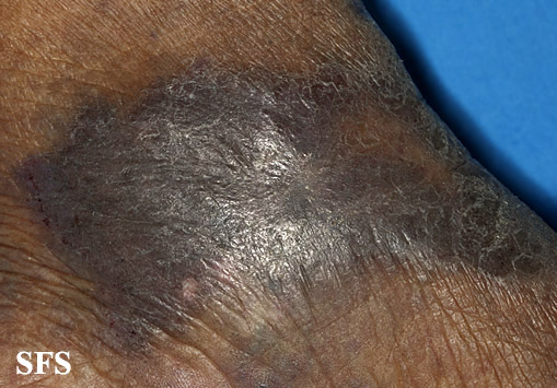
Acroangiodermatitis of Mali (also known as “Mali acroangiodermatitis” and “Pseudo-Kaposi’s sarcoma”[1]) is a rare cutaneous condition often characterized by purplish-blue to brown papules and plaques on the medial and lateral malleolus of both legs.[1]:1604[2]
Acroangiodermatitis is a rare skin condition characterised by hyperplasia of pre-existing vasculature due to venous hypertension from severe chronic venous stasis. It is associated with amputees, haemodialysis (HD) patients with arteriovenous (AV) shunts, and patients with paralysed legs, hepatitis C, chronic venous insufficiency or AV malformations (AVM). Patients present with itchy, painful, confluent, violaceous or brown-black macules, papules or plaques usually at the distal lower limbs. There may be ulceration and bleeding. The histologic features are capillary proliferation and perivascular inflammation involving eosinophils in the dermis with minimal epidermal changes. Management includes compression therapy, wound care and surgical correction of AVM. Dapsone combined with leg elevation and compression, and erythromycin for HD patients with AV fistulas have also been reported. The lesions may persist for years with complications like ulceration, bleeding and infection.
Differential Diagnosis
Acroangiodermatitis should be differentiated from other conditions presenting as purplish-blue to brown papules and plaques on extremities. The differentials include the following:
| Diseases | Etiology | Congenital | Acquired | Demography | Clinical manifestations | Lab findings | Gold standard diagnosis | Associated findings | |||||||||||
|---|---|---|---|---|---|---|---|---|---|---|---|---|---|---|---|---|---|---|---|
| Symptoms | Signs | CBC | LFT | ESR/CRP | Histopathology | ||||||||||||||
| Appearance | Fever | Bleeding | BP | Hepatosplenomegaly | Lymphadenopathy | Other | WBC | Hb | Plt | ||||||||||
| Bacillary angiomatosis [3] | – | + | Any age, usually between 20 -50 years | Solitary or multiple red, purple, flesh-colored, or colorless papules | ± | ± | Nl | – | – | Nl | Nl | Nl | Nl | Nl |
|
Clinical manifestation | |||
| Arteriovenous malformation [4] | + | – | Any age | Nl | – | + | Nl | – | – | Nl | Nl | Nl | Nl | Nl | NA | Imaging | |||
| Acroangiodermatitis[5] |
|
– | – | Any age, more in males | Purplish-blue to brown papules and plaques | – | – | Nl | – | – |
|
Nl | Nl | Nl | Nl | Nl |
|
Clinical manifesttations | |
| Angiosarcoma [6] | – | – | Adults, more in males | Enlarging bruise, a blue-black nodule, or an unhealed ulceration | – | – | Nl | – | – | – | Nl | ↓ | ↓ | Nl | Nl |
|
Biopsy | NA | |
| Diseases | Etiology | Congenital | Acquired | Demography | Appearance | Fever | Bleeding | BP | Hepatosplenomegaly | Lymphadenopathy | Other | WBC | Hb | Plt | LFT | ESR/CRP | Histopathology | Gold standard diagnosis | Associated findings |
| Masson’s hemangioma [7] | – | – | Rare |
|
– | – | Nl | – | – | – | Nl | Nl | Nl | Nl | Nl |
|
Biopsy | ||
| Seborrheic keratosis [8] |
|
+ | – | Any age |
|
– | – | Nl | – | – | – | Nl | Nl | Nl | Nl | Nl |
|
Clinical manifestations |
|
| Systemic lupus erythematosus (SLE) [9] | – | – | More common in female, typically in the 20 to 30 years |
|
± | – | ↑ | ± | ± | ↑ | ↓ | ↓ | Nl | Nl |
|
Clinical manifestations | |||
| Pyogenic granuloma [10] | + | + | Any age, usually in 20-30 years |
|
– | + | Nl | – | – | – | Nl | Nl | Nl | Nl | Nl |
|
Clinical manifestation | NA | |
| Benign lymphangioendothelioma [11] | – | + | Any ages, median age is 50 years | – | – | Nl | – | – | – | Nl | Nl | Nl | Nl | Nl |
|
Biopsy | NA | ||
| Cavernous hemangioma [12] | – | – | Usually in third to fifth decades of life. |
|
– | – | Nl | – | – | – | Nl | Nl | Nl | Nl | Nl |
|
Clinical manidestation |
| |
| Diseases | Etiology | Congenital | Acquired | Demography | Appearance | Fever | Bleeding | BP | Hepatosplenomegaly | Lymphadenopathy | Other | WBC | Hb | Plt | LFT | ESR/CRP | Histopathology | Gold standard diagnosis | Associated findings |
Physical examination
Gallery
-
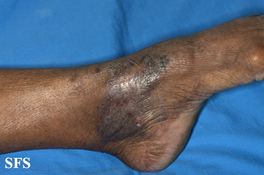 Acroangiodermatitis.
Adapted from [http://www.atlasdermatologico.com.br/disease.jsf?diseaseId=9 -
 Acroangiodermatitis.
Adapted from [http://www.atlasdermatologico.com.br/disease.jsf?diseaseId=9 -
 Acroangiodermatitis.
Adapted from [http://www.atlasdermatologico.com.br/disease.jsf?diseaseId=9


References
- ↑ 1.0 1.1 Rapini, Ronald P.; Bolognia, Jean L.; Jorizzo, Joseph L. (2007). Dermatology: 2-Volume Set. St. Louis: Mosby. ISBN 1-4160-2999-0.
- ↑ Scholz S, Schuller-Petrovic S, Kerl H (March 2005). “Mali acroangiodermatitis in homozygous activated protein C resistance”. Arch Dermatol. 141 (3): 396–7. doi:10.1001/archderm.141.3.396. PMID 15781687.
- ↑ Tappero JW, Perkins BA, Wenger JD, Berger TG (July 1995). “Cutaneous manifestations of opportunistic infections in patients infected with human immunodeficiency virus”. Clin. Microbiol. Rev. 8 (3): 440–50. PMC 174635. PMID 7553576.
- ↑ Whitehead KJ, Smith MC, Li DY (February 2013). “Arteriovenous malformations and other vascular malformation syndromes”. Cold Spring Harb Perspect Med. 3 (2): a006635. doi:10.1101/cshperspect.a006635. PMC 3552339. PMID 23125071.
- ↑ Lugović L, Pusić J, Situm M, Buljan M, Bulat V, Sebetić K, Soldo-Belić A (2007). “Acroangiodermatitis (pseudo-Kaposi sarcoma): three case reports”. Acta Dermatovenerol Croat. 15 (3): 152–7. PMID 17868541.
- ↑ Barttelbort SW, Stahl R, Ariyan S (July 1989). “Cutaneous angiosarcoma of the face and scalp”. Plast. Reconstr. Surg. 84 (1): 55–9. PMID 2734404.
- ↑ Park KK, Won YS, Yang JY, Choi CS, Han KY (July 2012). “Intravascular Papillary Endothelial Hyperplasia (Masson tumor) of the Skull : Case Report and Literature Review”. J Korean Neurosurg Soc. 52 (1): 52–4. doi:10.3340/jkns.2012.52.1.52. PMC 3440504. PMID 22993679.
- ↑ Noiles K, Vender R (2008). “Are all seborrheic keratoses benign? Review of the typical lesion and its variants”. J Cutan Med Surg. 12 (5): 203–10. doi:10.2310/7750.2008.07096. PMID 18845088.
- ↑ Uva L, Miguel D, Pinheiro C, Freitas JP, Marques Gomes M, Filipe P (2012). “Cutaneous manifestations of systemic lupus erythematosus”. Autoimmune Dis. 2012: 834291. doi:10.1155/2012/834291. PMC 3410306. PMID 22888407.
- ↑ Kamal R, Dahiya P, Puri A (January 2012). “Oral pyogenic granuloma: Various concepts of etiopathogenesis”. J Oral Maxillofac Pathol. 16 (1): 79–82. doi:10.4103/0973-029X.92978. PMC 3303528. PMID 22434943.
- ↑ Guillou L, Fletcher CD (August 2000). “Benign lymphangioendothelioma (acquired progressive lymphangioma): a lesion not to be confused with well-differentiated angiosarcoma and patch stage Kaposi’s sarcoma: clinicopathologic analysis of a series”. Am. J. Surg. Pathol. 24 (8): 1047–57. PMID 10935645.
- ↑ Goldberg RE, Pheasant TR, Shields JA (December 1979). “Cavernous hemangioma of the retina. A four-generation pedigree with neurocutaneous manifestations and an example of bilateral retinal involvement”. Arch. Ophthalmol. 97 (12): 2321–4. PMID 229814.
© 2026 MyEClinic – IFTM Institut für Telematik in der Medizin GmbH