
Anatomy of the heart external and internal features of the heart
Editor-In-Chief: C. Michael Gibson, M.S., M.D. [1]; Assistant Editor(s)-in-Chief: Rim Halaby, Yazan Daaboul
External and Internal Features of the Heart
Layers of the Heart
- The heart wall consists of 3 layers:
- Epicardium: thin external layer formed by visceral layer of serous pericardium
- Myocardium: thick middle layer made of cardiac muscle
- Endocardium: thin internal membrane that lines the heart and its valves. It is composed of endothelium and sub-endothelial connective tissue, similar to intimal component of blood vessels.
External Features
- The heart and roots of great vessels are embedded in the pericardial sac, approximately in the center of the thorax. The size of the heart is slightly larger than a clenched fist. The heart is surrounded laterally and posteriorly by the lungs, and anteriorly bound by the sternum and medial sections of the ribs and sterno-costal joints.
- It has the shape of a three-sided pyramid with an apex (left anterior), a base (posterior), and 4 surfaces: Sterno-costal/anterior (formed by right ventricle), diaphragmatic/inferior (formed by left ventricle and part of right ventricle), left pulmonary (formed by left ventricle, in contact with left lung), and right pulmonary (formed by right atrium).
- The heart appears trapezoid in the posterior and anterior views. As such, it consists of 4 borders: Right (convex), Inferior (horizontal), left (oblique), and superior.
- Below is an image showing a schematic outline of the heart in the mediastinum.

- The external surface of the heart is notable for 3 main sulci (grooves):
- Coronary (atrioventricular) sulcus: runs around the heart, and separates atria from ventricles
- Anterior interventricular sulcus: runs along the interventricular septum anteriorly
- Posterior interventricular sulcus: runs along the interventricular septum posteriorly
Internal Features
- The heart has 4 chambers: Right and left atria and right and left ventricles.
- Blood flows normally in the right to left direction: Right atrium to right ventricle to left atrium, and finally to left ventricle.
- Oxygen-depleted blood reaches the right atrium via the coronary sinus and superior and inferior vena cava and evacuates the right ventricle via the pulmonary artery towards the lungs.
- Oxygen enriched blood then re-enters the left atrium via 4 pulmonary veins and evacuates the left ventricle via the aorta.
- Below is an image showing the atria and ventricles of the heart as well as arrows indicating the normal blood flow throught the heart valves.
-
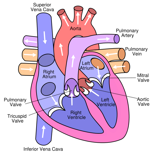 Anterior (frontal) view of the opened heart. White arrows indicate normal blood flow.

References
© 2026 MyEClinic – IFTM Institut für Telematik in der Medizin GmbH