
Asherman's syndrome other imaging findings

Editor-In-Chief: C. Michael Gibson, M.S., M.D. [1] Associate Editor-In-Chief: Saud Khan M.D.
Overview
Saline sonography or hysterosalpingography may be used initially in the evaluation of Asherman’s syndrome. Though hysteroscopy remains the gold standard.
Diagnosis
Hysteroscopy is the gold standard for diagnosis [1]. Imaging by sonohysterography or hysterosalpingography will reveal the extent of the scar formation. Hormone studies show normal levels consistent with reproductive function. Advantages of saline sonography compared with hysterosalpingography are that it does not involve radiation or a special suite, it may be done in-office. Hysterosalpingography must be performed in a radiology suite. The advantage of Hysterosalpingography is the feature that allows evaluation of tubal patency, although newer techniques for saline sonography that involve infusion of a water/air combination may improve assessment of tubal patency.[2]
Hysterosalpingography
(Images courtesy of RadsWiki)
-
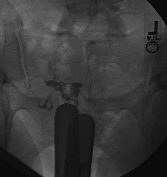 Hysterosalpingography: Asherman’s syndrome
 -
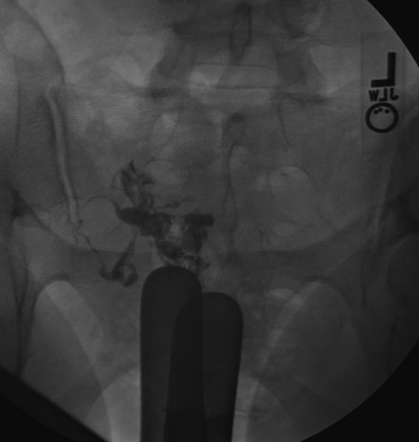 Hysterosalpingography: Asherman’s syndrome
 -
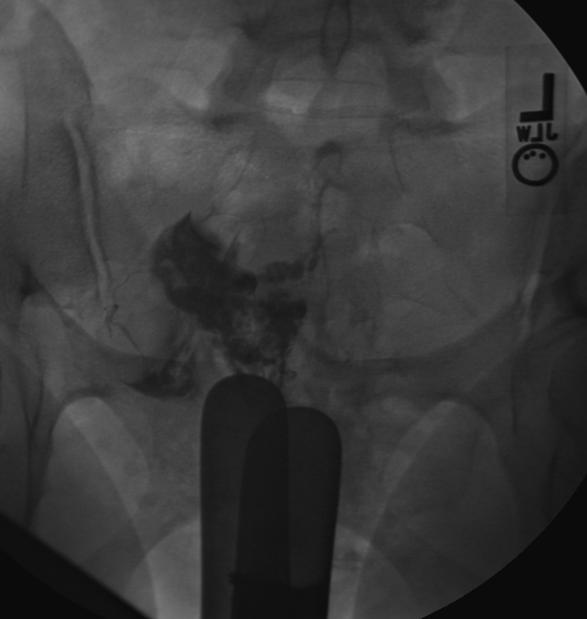 Hysterosalpingography: Asherman’s syndrome
 -
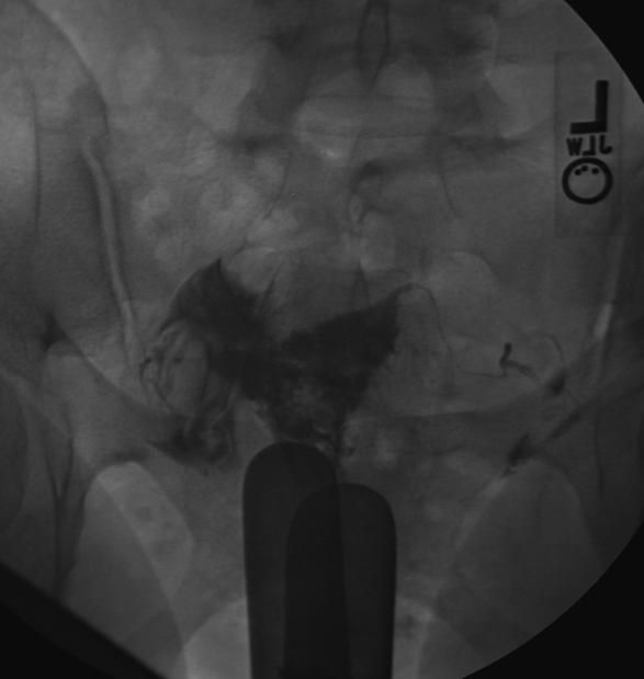 Hysterosalpingography: Asherman’s syndrome


References
- ↑ Valle RF, Sciarra JJ. Intrauterine adhesions: hysteroscopic diagnosis, classification, treatment, and reproductive outcome. Am J Obstet Gynecol 1988; 158:1459-1470.
- ↑ Luciano DE, Exacoustos C, Johns DA, Luciano AA (2011). “Can hysterosalpingo-contrast sonography replace hysterosalpingography in confirming tubal blockage after hysteroscopic sterilization and in the evaluation of the uterus and tubes in infertile patients?”. Am J Obstet Gynecol. 204 (1): 79.e1–5. doi:10.1016/j.ajog.2010.08.065. PMID 21187197.
© 2026 MyEClinic – IFTM Institut für Telematik in der Medizin GmbH