
Atelectasis chest x ray
Editor-In-Chief: C. Michael Gibson, M.S., M.D. [1]; Associate Editor(s)-in-Chief: Sudarshana Datta, MD [2]
Overview
An x-ray may be helpful in the diagnosis of atelectasis. Findings on an x-ray suggestive of atelectasis include displacement of fissures, rib crowding, elevation of ipsilateral diaphragm, volume loss on ipsilateral hemithorax, hilar displacement and compensatory hyperlucency of the remaining lobes. Complete lung atelectasis and atelectasis involving different parts of the lung have their own characteristic appearance. While complete atelectasis of the lung may lead to opacification of the entire hemithorax and ipsilateral shift of the mediastinum, a right midle and lower lobe atelectasis may show subpulmonic effusions along with right hemidiaphragmatic elevation on X-ray.
X Ray
- Atelectasis of the lung is a very common abnormality seen on chest radiographs. Abnormalities on chest X-ray due to atelectasis help in the delineation of the underlying pathology.[1]
- Different types of atelectasis have their own characteristic radiographic pattern and etiology.
- An x-ray may be helpful in the diagnosis of atelectasis. Findings on an x-ray suggestive of atelectasis include:[2]
- Signs of lobar collapse such as:
- Shifting of the mediastinum towards the collapsed lung lobe
- Hilar displacement
- Silhouetting of the diaphragm or the heart border
- Rib crowding
- Compensatory hyperlucency of the remaining lobes
- Elevation of ipsilateral diaphragm
- Opacification of the collapsed lung lobe
- Displacement of fissures
- Volume loss on ipsilateral hemithorax
- Air bronchograms help delineate the site of obstruction
- Signs of lobar collapse such as:
- X-ray findings in cases with complete atelectasis of the lung include:[3][4]
- Opacification of the entire hemithorax due to complete collapse of a lung
- Ipsilateral shift of the mediastinum, that helps distinguish atelectasis from pleural effusion
- X-ray findings suggestive of right upper lobe (RUL) collapse include:
- X-ray appearance of right middle lobe collapse:
- Triangular opacity
- X-ray appearance of right lower lobe (RLL) collapse:
- Posterior and inferior shift of RLL due to collapse
- Superior triangle sign: Rightward shift of structures in the superior mediastinum
- Blurring of the right hemidiaphragm (posterior third)
- Visibility of the major fissure, which is usually not seen
- X-ray appearance of a right middle and lower lobe atelectasis:[5]
- Subpulmonic effusion
- Elevation of the right hemidiaphragm
- X-ray appearance of left upper lobe (LUL) collapse:
- Atelectatic left upper lobe shifts anteriorly and superiorly
- PA view: Faint opacity of the atelectatic lobe in the left upper hemithorax
- X-ray appearance of left lower lobe (LLL) collapse:
- Retrocardiac opacity
- Downward displacement of the hilum
- Aortic-knob sign: Obliteration of the aortic arch by the superior mediastinum
- Lateral view: Indistinct appearance of the posterior third of the diaphragm due to opacity
- X-ray appearance of rounded atelectasis:
- Subpleural mass
- Location of rounded atelectasis: Right middle lobe, lower lobes or lingula
- Comet-tail sign or talon sign: Bronchovascular structures projecting out of the mass toward the hilum, in a swirl appearance
- Parietal pleural plaque
- X-ray appearance of post-surgical atelectasis:
- Bibasal pattern
- CXR also helps determine the efficacy of chest physiotherapy in patients with atelectasis.
Images shown in this section are courtesy of RadsWiki and copylefted.
-
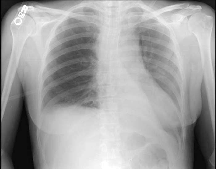
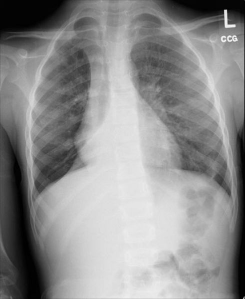 Right lower lobe collapse
 -
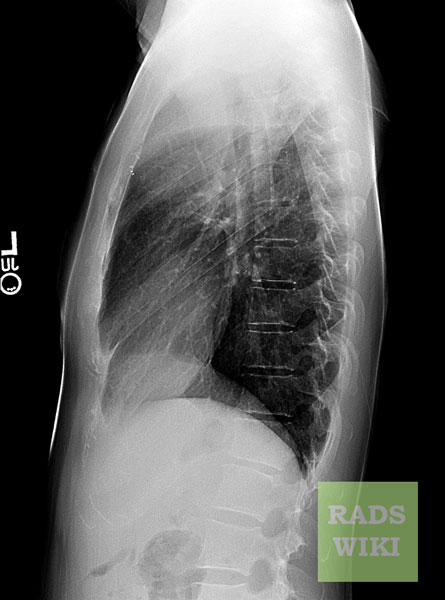
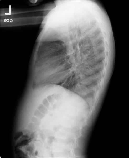 Right lower lobe collapse. The same patient. Lateral view.


-
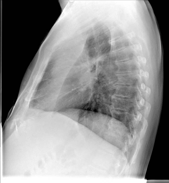
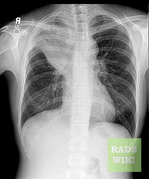
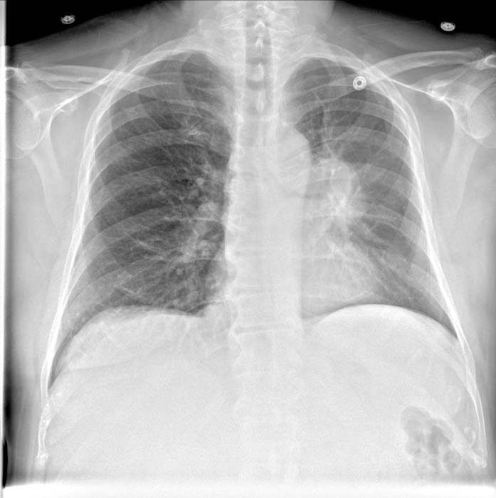 Left upper lobe collapse
 -
 Left upper lobe collapse


-
 Left lower lobe collapse

-
 S Sign of the Golden
 -
 S Sign of the Golden


References
- ↑ Ashizawa K, Hayashi K, Aso N, Minami K (January 2001). “Lobar atelectasis: diagnostic pitfalls on chest radiography”. Br J Radiol. 74 (877): 89–97. doi:10.1259/bjr.74.877.740089. PMID 11227785.
- ↑ Qureshi NR, Gleeson FV (June 2006). “Imaging of pleural disease”. Clin. Chest Med. 27 (2): 193–213. doi:10.1016/j.ccm.2006.02.001. PMID 16716813.
- ↑ Woodring JH, Reed JC (1996). “Radiographic manifestations of lobar atelectasis”. J Thorac Imaging. 11 (2): 109–44. PMID 8820022.
- ↑ Proto AV, Tocino I (April 1980). “Radiographic manifestations of lobar collapse”. Semin Roentgenol. 15 (2): 117–73. PMID 7394541.
- ↑ Stark P, Leung A (1996). “Effects of lobar atelectasis on the distribution of pleural effusion and pneumothorax”. J Thorac Imaging. 11 (2): 145–9. PMID 8820023.
© 2026 MyEClinic – IFTM Institut für Telematik in der Medizin GmbH