
Bile duct cyst pathophysiology
Editor-In-Chief: C. Michael Gibson, M.S., M.D. [1]
Overview
Pathophysiology
Bile Duct Anatomy
The path is as follows: Bile canaliculi → Canals of Hering → interlobular bile ducts → intrahepatic bile ducts → left and right hepatic ducts merge to form → common hepatic duct exits liver and joins → cystic duct (from gall bladder) forming → common bile duct → joins with pancreatic duct → forming ampulla of Vater → enters duodenum
-
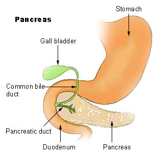 Region of pancreas
 -
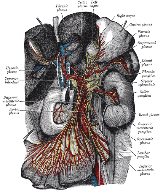 The celiac ganglia with the sympathetic plexuses of the abdominal viscera radiating from the ganglia.
 -
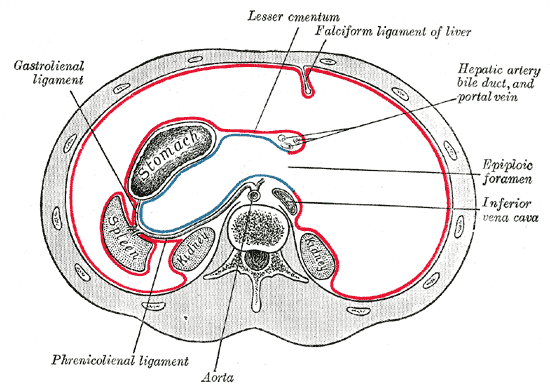 Horizontal disposition of the peritoneum in the upper part of the abdomen.
 -
 Interior of the descending portion of the duodenum, showing bile papilla.
 -
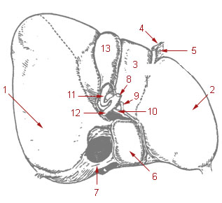
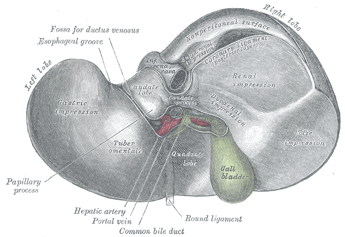 Inferior surface of the liver.
 -
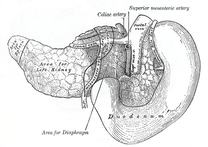 The pancreas and duodenum from behind.
 -
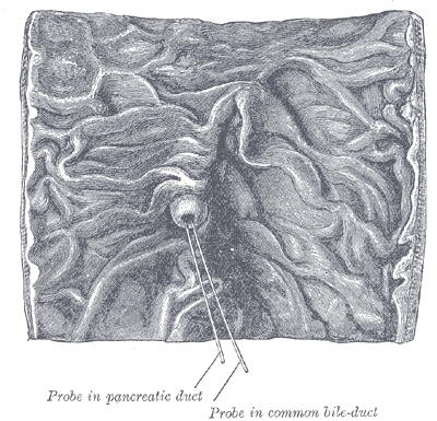
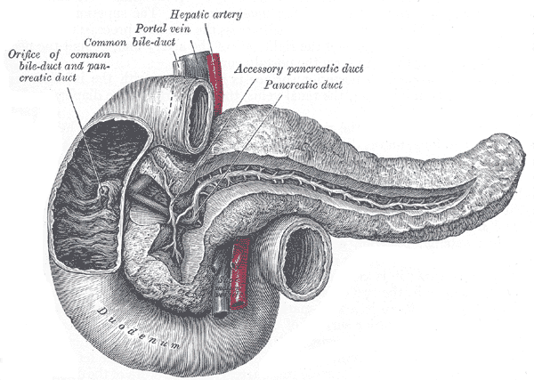 The pancreatic duct.
 -
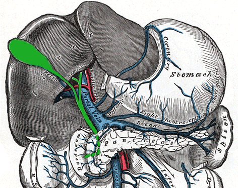 The portal vein and its tributaries.
 -
 Liver and gallbladder


References
© 2026 MyEClinic – IFTM Institut für Telematik in der Medizin GmbH