
Boerhaave syndrome chest x ray
Editor-In-Chief: C. Michael Gibson, M.S., M.D. [1] Associate Editor(s)-in-Chief: Mohamed Diab, MD [2], Shaghayegh Habibi, M.D.[3], Ajay Gade MD[4]], Feham Tariq, MD [5]
Overview
An upright anterior-posterior view of the chest is the most useful in early diagnosis. In most patients with Boerhaave syndrome (BHS) chest x-ray shows one-sided effusion, pneumothorax, hydropneumothorax, pneumomediastinum and subcutaneous emphysema.
Chest X Ray
- An upright anterior-posterior view of the chest is the most useful in early diagnosis, as most of the patients will reveal an abnormal chest finding after the perforation.
- The Naclerio V-sign may be seen on chest radiograph as radiolucent streaks of air seen in the retro-cardiac region in the V shape.[1]
The usual although unspecific radiographic features of BHS include:[2]
- One-sided effusion (usually on the left)
- Lung infiltrates
- Atelectasis
Whereas more specific signs are rarely detected or very subtle:[1][3][2]
- Pneumothorax
- Hydropneumothorax
- Pneumomediastinum
- Mediastinal widening
- Pneumopericardium
- Subcutaneous emphysema
-
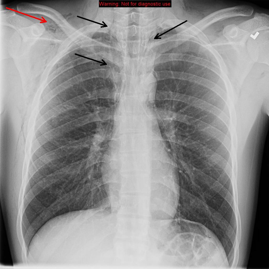 Chest X-ray: Frontal view reveals pneumomediastinum (black arrows). Subcutaneous emphysema (red arrow) along the chest wall, more prominent along the right than left; Source- Radiopaedia
 -
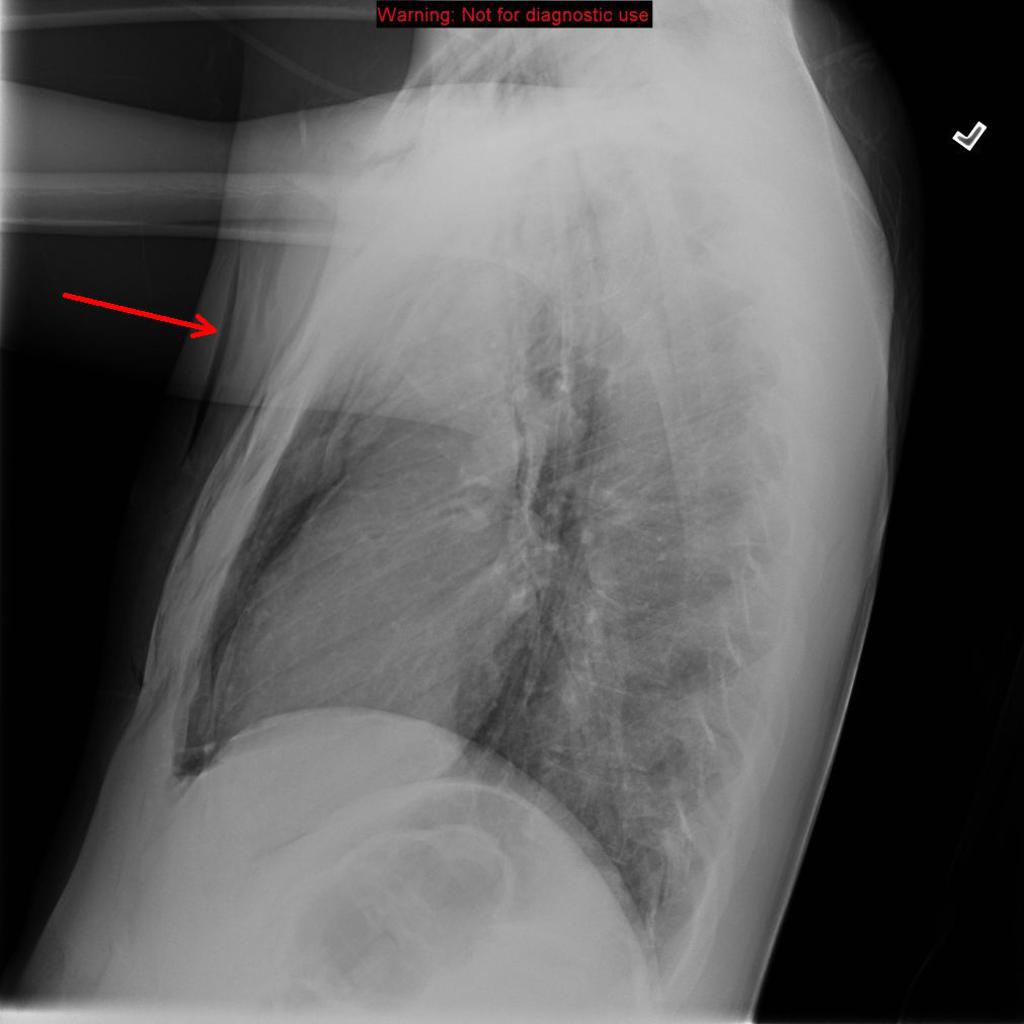 Chest X-ray: Boerhaave syndrome- Lateral radiographs subcutaneous emphysema (red arrow) along the chest wall; Source- Radiopaedia


References
- ↑ 1.0 1.1 Maurya VK, Sharma P, Ravikumar R, Bhatia M (2016). “Boerhaave’s syndrome”. Med J Armed Forces India. 72 (Suppl 1): S105–S107. doi:10.1016/j.mjafi.2015.12.004. PMC 5192176. PMID 28050085.
- ↑ 2.0 2.1 Tonolini M, Bianco R (2013). “Spontaneous esophageal perforation (Boerhaave syndrome): Diagnosis with CT-esophagography”. J Emerg Trauma Shock. 6 (1): 58–60. doi:10.4103/0974-2700.106329. PMC 3589863. PMID 23493470.
- ↑ Pate JW, Walker WA, Cole FH, Owen EW, Johnson WH (1989). “Spontaneous rupture of the esophagus: a 30-year experience”. Ann. Thorac. Surg. 47 (5): 689–92. PMID 2730190.
© 2026 MyEClinic – IFTM Institut für Telematik in der Medizin GmbH