
Cavernous angioma MRI
Editor-In-Chief: C. Michael Gibson, M.S., M.D. [1]<nowiki>; Associate Editor(s)-in-Chief: Edzel Lorraine Co, D.M.D., M.D.
Overview
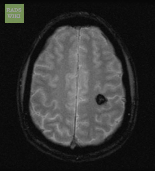
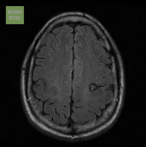
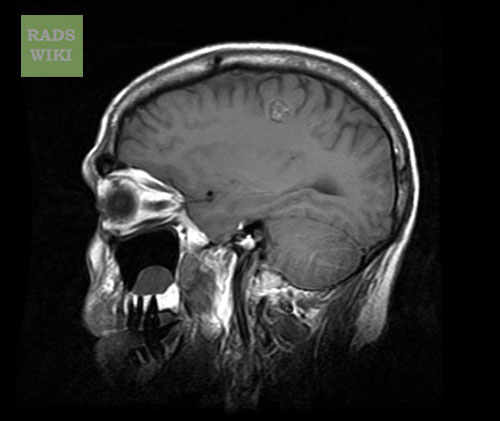
Diagnosis can be made through incidental findings from magnetic resonance imaging (MRI) screening. A gradient-echo sequence should be utilized to unmask punctate lesions which can go undetected. These lesions are more visible on FLAIR imaging than on T2 weighing. As compared to T2 weighing, FLAIR imaging has more suppression of free-flowing fluid signals.
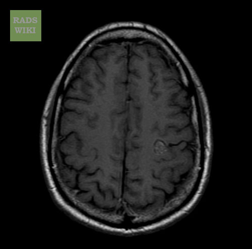
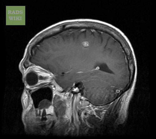
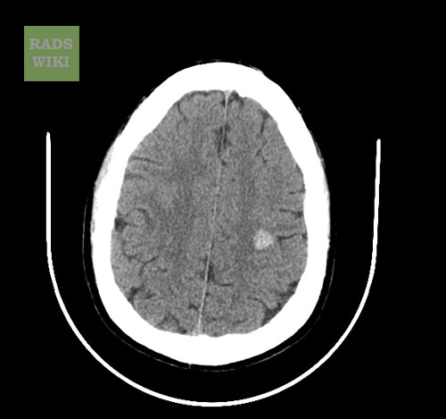
- These lesions are usually described as popcorn-like, smooth, well-circumscribed complex lesions.[1]
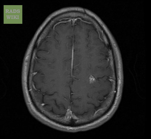
- There is a foci of mixed-signal intensities in the core, which signifies the presence of hemorrhage in different stages of evolution.
- On T1-weighted images, there is a low-signal-intensity hemosiderin rim bordering the heterogeneous core.
- Focal hypointense nodules are usually associated with smaller cavernous malformation lesions in both T1- and T2-weighted sequences. These small lesions become more evident in gradient-echo images due to increased susceptibility effects of the sequences.
-
 MRI: Cavernous malformation
 -
 MRI: Cavernous malformation


-
 MRI: Cavernous malformation
 -
 MRI: Cavernous malformation


-
 MRI: Cavernous malformation
 -
 MRI: Cavernous malformation
 -
 MRI: Cavernous malformation


References
- ↑ Rafee S, Killeen RP, Tubridy N (2021). “‘Popcorn’ in the Brain: A Cause for Confusion”. Am J Med. 134 (2): 216–217. doi:10.1016/j.amjmed.2020.09.014. PMID 33091393 Check
|pmid=value (help).
© 2026 MyEClinic – IFTM Institut für Telematik in der Medizin GmbH