
Chorionic villi
Editor-In-Chief: C. Michael Gibson, M.S., M.D. [1]
Overview
Chorionic villi are villi that sprout from the chorion in order to give a maximum area of contact with the maternal blood.
Embryonic blood is carried to the villi by the branches of the umbilical arteries, and after circulating through the capillaries of the villi, is returned to the embryo by the umbilical veins.
Thus, the villi are part of the border between maternal and fetal blood during pregnancy.
Development
The chorion undergoes rapid proliferation and forms numerous processes, the chorionic villi, which invade and destroy the uterine decidua and at the same time absorb from it nutritive materials for the growth of the embryo.
They undergo several stages, depending on their composition.
| Stage | Description | Contents |
| Primary | The chorionic villi are at first small and non-vascular. | trophoblast only [1] |
| Secondary | The villi increase in size and ramify, while the mesoderm grows into them. | trophoblast and mesoderm [1] |
| Tertiary | Branches of the umbilical vessels, grows into the mesoderm, and in this way the chorionic villi are vascularized. | trophoblast, mesoderm, and blood vessels [1] |
Until about the end of the second month of pregnancy the villi cover the entire chorion, and are almost uniform in size, but after this they develop unequally.
Relations
The villi can also be classified by their relations:
Floating villi
These villi are found floating freely in the intervillous space. They exhibit a bi-layered epithelium consisting of cytotrophoblasts with overlaying syncytium (syncytiotrophoblast).
Anchoring (stem) villi
These villi act to stablise mechanical integrity of the placental-maternal interface.
Tissue composition and cell types
The bulk of the villi consist of connective tissues in which blood vessels are found. Most of the cells in the connective tissue core of the villi are fibroblasts. Macrophages known as Hofbauer cells are also present.
Additional images
-
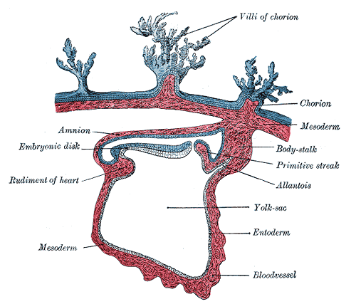 Section through the embryo.
 -
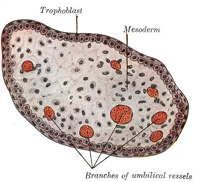 Transverse section of a chorionic villus.
 -
Primary chorionic villi. Diagrammatic.
-
Secondary chorionic villi. Diagrammatic.
-
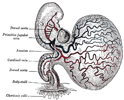 Human embryo of about fourteen days, with yolk-sac.


See also
References
© 2026 MyEClinic – IFTM Institut für Telematik in der Medizin GmbH