
EKG abnormalities in central nervous system disease
Editor-In-Chief: C. Michael Gibson, M.S., M.D. [1]; Associate Editor-In-Chief: Cafer Zorkun, M.D., Ph.D. [2]
Overview
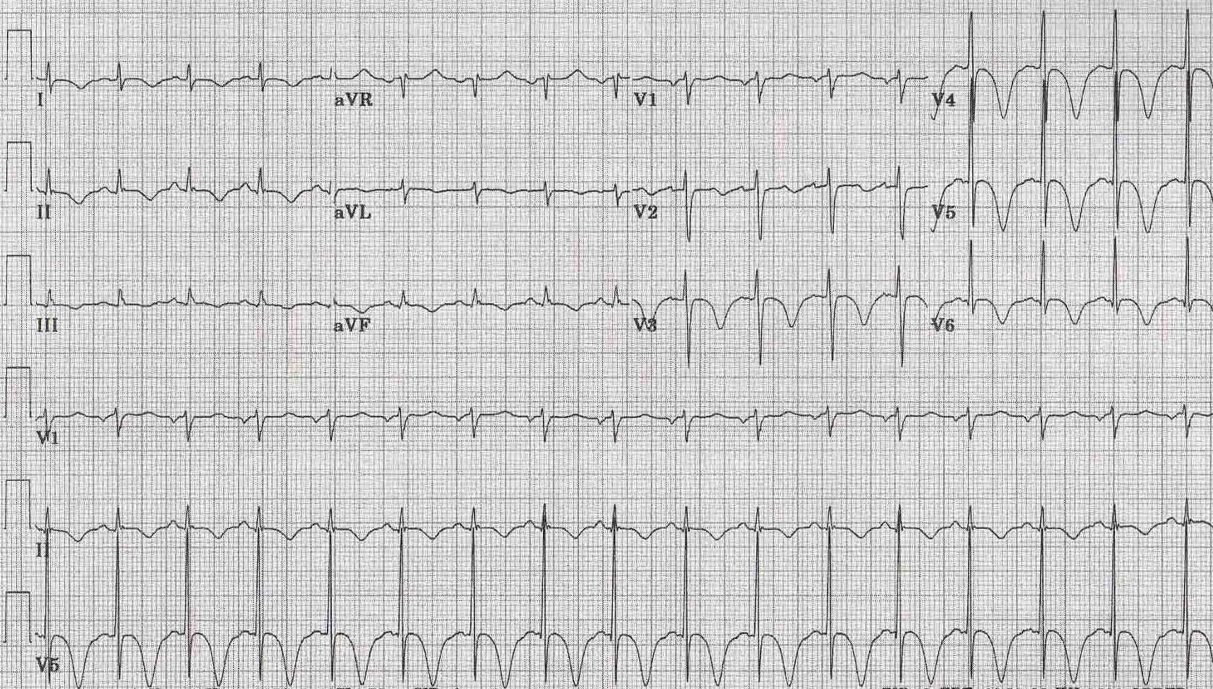
Classic manifestations on EKG of the so-called cerebrovascular accidents, (most commonly associated with subarachnoid hemorrhage or other intracranial bleeds) are symmetrically and deeply inverted giant T waves.
Pathophysiology
The mechanisms are not fully delineated but may relate to excessive catecholamine stimulation causing direct myocardial injury (myocytolysis).
Diagnosis
Electrocardiographic Findings
The ECG may be notable for marked QT-U prolongation (sometimes a giant U wave appears to be embeded in the T wave, creating a slight discontinuity in the waveform morphology). The long QT-U may predispose to torsade(s) de pointes. Takostubo syndrome may occur in some cases. Apical hypertrophic cardiomyopathy (Yamaguchi’s syndrome) is associated with deep narrow (spade-like) T wave inversions, most marked in the mid-precordial leads.
- EKG changes seen in 71.5% of patients with subarachnoid hemorrhage, and 57.1% of those with cerebral hemorrhage.
- Most common abnormalities are
- Large, upright, or deeply inverted T waves
- Prolongation of the QTc interval
- Prominent U waves
- Can persist for 11 days
- Rarely can ST segment elevation or depression
- Rhythm disturbances
- Reason for changes is thought to be altered autonomic tone
Examples
-
 Patient with subaracnoidal hemorrhage
 -
 EKG of a patients with CNS Disorders
 -
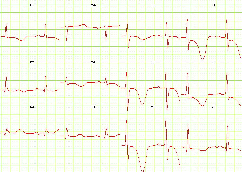 Inverted and deep T waves in the precordal leads due to subaracnoidal aneurysm [Image courtesy of Dr Jose Ganseman Dr Ganseman’s webpage: An ultimate source of EKG


Refrences
© 2026 MyEClinic – IFTM Institut für Telematik in der Medizin GmbH