
Echo in Ebstein's anomaly of the tricuspid valve
Editors: Eli V. Gelfand, MD and Keri Shafer, MD (Beth Israel Deaconess Medical Center, Harvard Medical School, Boston, MA)
Ebstein’s Anomaly is the apical displacement of the tricuspid valve into the right ventricle. For more see: Ebstein’s Anomaly of the Tricuspid Valve
-
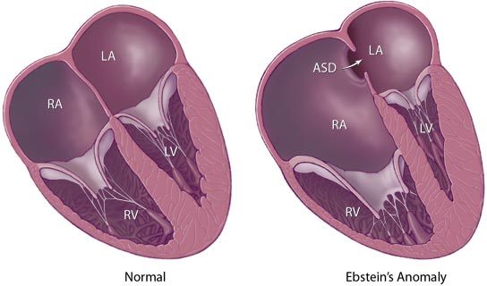 Graphical represntation of Ebstein’s Anomaly from the Mayo Clinic website (note there is also an ASD on this diagram)

Echo functions in Ebstein’s Anomaly
Echocardiographic evaluation of Ebstein’s must include evaluation of:
- extent of apical displacement and attachment location of the leaflets
- degree of tricuspid regurgitation
- morphology of the anterior leaflet
- presence or absence of right ventricular outflow tract obstruction
- degree of atrialization
- surgical options
- presence of mitral valve prolapse
- associated anomalities including Atrial Septal Defect and less commonly Ventricular Septal Defect
Special Echo techniques in Ebstein’s Anomaly
- Measurement of the distance between the insertion sites of the tricuspid leaflets
- Doppler should be used to determine the degree of tricuspid regurgitation
Ebstein’s Anomaly is best seen in the apical four chamber view
Echocardiographic representation from in various views
Apical 4-chamber:

Apical continuous-wave doppler:

Short axis view (notice the tethering of the tricuspid valve):

Below in a video clip is an apical 4-chamber transthoracic view, showing apically-displaced leaflets of the tricuspid valve. Note substantial tricuspid regurgitation, which originates close to the apex, where the abnormal leaflets actually coapt:
{{#ev:youtube|KEko3kM26bY}}
External Links
- Yale Congenital Heart Disease- Ebstein’s Anomaly
- Emedicine Ebstein’s Anomaly
- Mayo Clinic Ebstein’s Anomaly
© 2026 MyEClinic – IFTM Institut für Telematik in der Medizin GmbH