
Electrocardiographic findings in right ventricular hypertrophy
Editor-In-Chief: C. Michael Gibson, M.S., M.D. [2]; Associate Editor-In-Chief: Cafer Zorkun, M.D., Ph.D. [3]
Overview
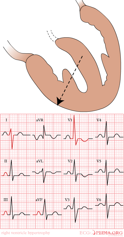
The general electrocardiographic findings of right ventricular hypertrophy include right axis deviation, an R/S ratio > 1 in V1, and the presence of P pulmonale.
Summary of EKG Criteria for RVH
- Right axis deviation of +90 degrees or more
- The R wave in V1 is 7 mm or more in height
- RV1 + SV5 or SV6 = 10 mm or more
- R/S ratio in V1 = 1.0 or more
- S/R ratio in V6 = 1.0 or more
- Late intrinsicoid deflection in V1 (0.035+)
- Incomplete RBBB pattern
- ST T strain pattern in 2,3,aVF
- P pulmonale or P congenitale
- S1 S2 S3 pattern in children
Differential Diagnosis of R>S in V1
- RVH
- Posterior MI
- WPW
- HCM (septal hypertrophy)
- Kulbertus’ block (septal fascicular block)
- Duchennes Muscular Dystrophy
- Normal variant
- V4r may be a more useful and reliable than lead V1 in that it often reveals an r>s while v1 remains normal
- An incomplete right bundle branch block in the right precordial chest leads may signal the development of RVH
- In the limb leads right axis deviation develops and at times prominent Q waves simulating an IMI appear in leads 2,3, and aVF.
- In children an S1 S2 S3 pattern (i.e. an S wave deeper than R in all 3 standard leads) is a reliable index of RVH
- RV strain can be seen in leads V1 and V2 but also in leads 2,3, aVF
Electrocardiographic Examples of RVH
-
 Right ventricular hypertrophy
 -
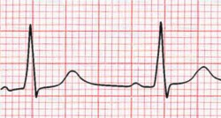 Right ventricular hypertrohpy, the R wave > the S wave in V1


© 2026 MyEClinic – IFTM Institut für Telematik in der Medizin GmbH