
Internal thoracic artery
Editor-In-Chief: C. Michael Gibson, M.S., M.D. [1]
Overview
In human anatomy, the internal thoracic artery (ITA), previously known as the internal mammary artery (a name still common among surgeons), is an artery that supplies the anterior chest wall and the breasts. It is a paired artery, with one running on each side of the body.
Course
The internal thoracic artery arises from the subclavian artery near its origin.
It travels downward on the inside of the ribcage, approximately a centimeter from the sides of the sternum, and thus medial to the nipple.
It runs posterior to the internal intercostal muscles, but anterior to the transverse thoracic muscles.
It continues downward until it divides into the musculophrenic artery and the superior epigastric artery around the sixth intercostal space.
Branches
- Mediastinal branches
- Thymic branches
- Pericardiophrenic artery – travels with the phrenic nerve
- Sternal branches
- Perforating branches
- Twelve anterior intercostal branches, two to each of the top six intercostal spaces. In a given space, the upper branch travelling laterally along the botton of the rib until it anastomoses with its corresponding posterior intercostal artery. The lower branch of the space anastomoses with a collateral branch of the posterior intercostal artery.
After passing the sixth intercostal space, the internal thoracic artery splits into the following two terminal branches:
- Musculophrenic artery – roughly follows the costal margin
- Superior epigastric artery – continues the course of the internal thoracic artery, travelling downward into the abdominal wall
Revascularization with the ITA
The internal thoracic artery is the cardiac surgeon’s blood vessel of choice for coronary artery bypass grafting. The left ITA has a superior long-term patency to saphenous vein grafts[1][2] and other arterial grafts[3] (e.g. radial artery, gastroepiploic artery) when grafted to the left anterior descending coronary artery, generally the most important vessel, clinically, to revascularize.
Diagnostic Cardiac Catheterization of the Left Internal Mammary Artery
In order to engage the left internal mammary artery, a view with slight cranial, and slight left anterior oblique (LAO) angulation is used. a Judkin’s Right 4 catheter (JR 4) catheter is used to engage the ostium of the subclavian artery. With the catheter near the anticipated origin of the subclavian artery, a counterclockwise motion of the catheter is needed to engage the ostium. Once the ostium is engaged, an exchange length 0.038 J tipped wire is then advance into the subclavian artery. A left internal mammary artery (LIMA) catheter is then exchanged to engage the origin of the left internal mammary artery. Again, this engagement is accomplished with counterclockwise motion of the catheter. An injection with the camera in this angulation is a suitable first view of the LIMA, with careful attention in this view paid to the ostium of the LIMA. the next view is often a left anterior oblique (LAO) view with 90 degrees of angulation (LAO 90). This allows for accurate assessment of the LIMA anastomosis to the left anterior descending artery.
Additional images
-
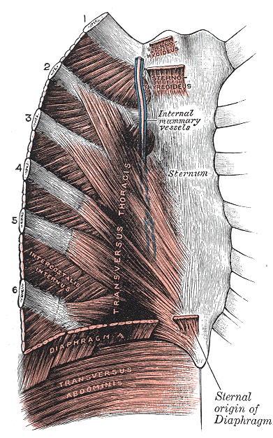 Posterior surface of sternum and costal cartilages, showing Transversus thoracis.
 -
 Transverse section of thorax, showing relations of pulmonary artery.
 -
 The axillary artery and its branches.
 -
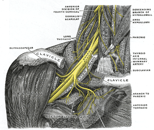 The right brachial plexus with its short branches, viewed from in front.
 -
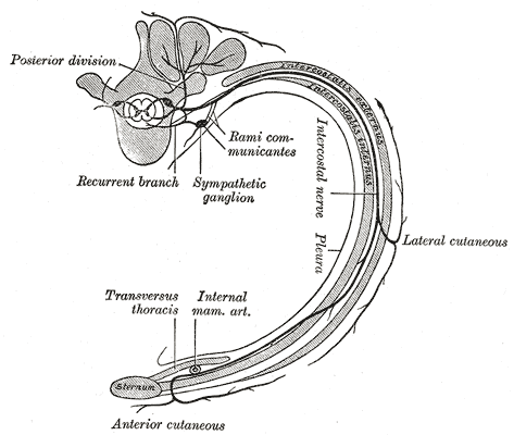 Diagram of the course and branches of a typica intercostal nerve.


References
- ↑ Kitamura S, Kawachi K, Kawata T, Kobayashi S, Mizuguchi K, Kameda Y, Nishioka H, Hamada Y, Yoshida Y. [Ten-year survival and cardiac event-free rates in Japanese patients with the left anterior descending artery revascularized with internal thoracic artery or saphenous vein graft: a comparative study] Nippon Geka Gakkai Zasshi. 1996 Mar;97(3):202-9. PMID 8649330.
- ↑ Arima M, Kanoh T, Suzuki T, Kuremoto K, Tanimoto K, Oigawa T, Matsuda S. Serial Angiographic Follow-up Beyond 10 Years After Coronary Artery Bypass Grafting. Circ J. 2005 Aug;69(8):896-902. PMID 16041156. Free Full Text.
- ↑ Cohen G, Tamariz MG, Sever JY, Liaghati N, Guru V, Christakis GT, Bhatnagar G, Cutrara C, Abouzahr L, Goldman BS, Fremes SE. The radial artery versus the saphenous vein graft in contemporary CABG: a case-matched study. Ann Thorac Surg. 2001 Jan;71(1):180-5; discussion 185-6. PMID 11216742.
External links
- Template:GPnotebook – Internal thoracic artery
Figures of ITA grafts
- Figure of heart with two saphenous vein grafts (SVGs) and a LITA graft – texheartsurgeons.com
- Drawing of the heart with a SVG to the right coronary artery (RCA) and a LITA graft to the LAD – darcystudios.com
- Drawing of the heart with a SVG to the RCA and a LITA graft to the LAD – mayoclinic.org
© 2026 MyEClinic – IFTM Institut für Telematik in der Medizin GmbH