
Intracerebral hemorrhage MRI
Editor-In-Chief: C. Michael Gibson, M.S., M.D. [1]; Associate Editor(s)-in-Chief: Sara Mehrsefat, M.D. [2]
Overview
MRI is better than CT for detection of acute and chronic hemorrhage. Therefore it should be the preferred test for accurate diagnosis of patients with suspected intracerebral hemorrhage.[1]
MRI
MRI is better than CT for detection of acute and chronic hemorrhage. Therefore it should be the preferred test for accurate diagnosis of patients with suspected intracerebral hemorrhage.[1]
- T2 susceptibility-weighted MRI are as sensitive as CT for detection of acute blood and are more sensitive for identification of prior hemorrhage.[1]
- The appearance of Intracerebral hemorrhage on MRI depends on the age of the hematoma and the type of MR sequence (T1-MRI or T2-MRI).
- As the hematoma ages, hemoglobin (Hb) goes through the following stages:[2]
| Stage | Age | Hemoglobin | 1-MRI | 2-MRI |
|---|---|---|---|---|
| Hyperacute | < 24 hours | Intracellular oxy-Hb | Isointense | Very hyperintense |
| Acute | 1-3 days | Intracellular deoxy-Hb | Slightly hypointense | Very hypointense |
| Early subacute | > 3 days | Intracellular met-Hb | Very hyperintense | Very hypointense |
| Late subacute | > 7 days | Extracellular met-Hb | Very hyperintense | Very hypointense |
| Chronic center | > 14 days | Extracellular hemichromes | Isointense | Very hyperintense |
| Chronic rim | > 14 days | Intracellular hemosiderin | Slightly hypointense | Very hypointense |
Microbleeds
Cerebral microbleeds are best visualized on T2* weighted GRE sequences as punctate areas of low signal.[3][4]
Images
The following are images associated with intracerebral hemorrhage involves the left lentiform nucleus and internal capsule.[5]
-
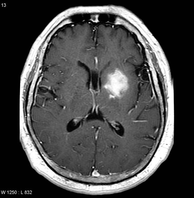 Axial T1
 -
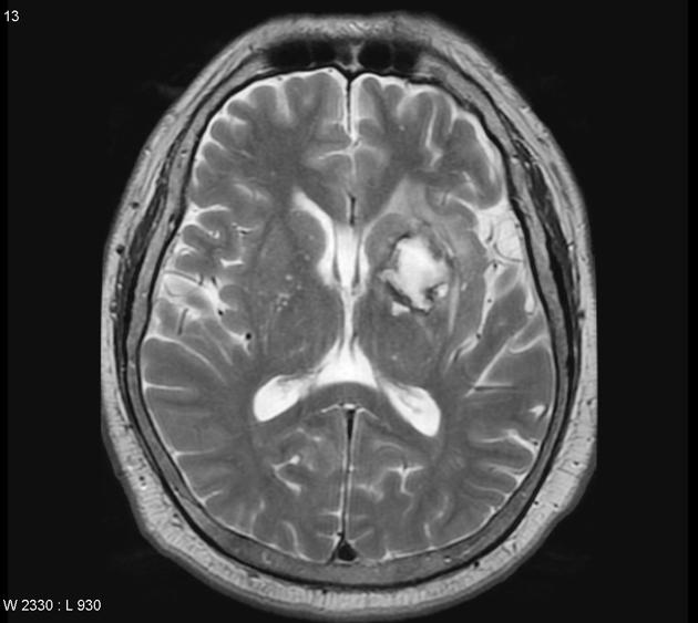 T2
T2 -
 Sagittal T1 C+


References
- ↑ 1.0 1.1 1.2 Chalela JA, Kidwell CS, Nentwich LM, Luby M, Butman JA, Demchuk AM; et al. (2007). “Magnetic resonance imaging and computed tomography in emergency assessment of patients with suspected acute stroke: a prospective comparison”. Lancet. 369 (9558): 293–8. doi:10.1016/S0140-6736(07)60151-2. PMC 1859855. PMID 17258669.
- ↑ Bradley WG (1993). “MR appearance of hemorrhage in the brain”. Radiology. 189 (1): 15–26. doi:10.1148/radiology.189.1.8372185. PMID 8372185.
- ↑ Altmann-Schneider I, Trompet S, de Craen AJ, van Es AC, Jukema JW, Stott DJ; et al. (2011). “Cerebral microbleeds are predictive of mortality in the elderly”. Stroke. 42 (3): 638–44. doi:10.1161/STROKEAHA.110.595611. PMID 21233474.
- ↑ Liu W, Liu R, Sun W, Peng Q, Zhang W, Xu E; et al. (2012). “Different impacts of blood pressure variability on the progression of cerebral microbleeds and white matter lesions”. Stroke. 43 (11): 2916–22. doi:10.1161/STROKEAHA.112.658369. PMID 22949472.
- ↑ Intracerebral Hemotrrhage https://radiopaedia.org/cases/intracerebral-haemorrhage-2 Accessed on November 9, 2016
© 2026 MyEClinic – IFTM Institut für Telematik in der Medizin GmbH