
Electrocardiographic findings in left ventricular hypertrophy
Editor-In-Chief: C. Michael Gibson, M.S., M.D. [2]; Associate Editor-In-Chief: Cafer Zorkun, M.D., Ph.D. [3]
Synonyms and keywords: LVH; LVH with strain; strain pattern
Overview
Left ventricular hypertrophy is associated with increased QRS voltage on the EKG and a strain pattern or inverted checkmark pattern to the T wave in the lateral leads. There are a variety of criteria used to diagnose left ventricular hypertrophy (LVH) on the EKG.
Diagnosis
Electrocardiography
Sokolow and Lyon Criteria
- Add the depth of the S wave in V1 to the height of the R wave in lead V5 or V6 (whichever is taller) and if the sum is greater than 35 mm then LVH is present.
- This criterion correlates well with the thickness of the LV walls and the diameter of the LV cavity as determined by ECHO.
- Sensitivity 22% and specificity of 100%.[1]
Effects of Left Anterior Hemiblock on Diagnosing Acute myocardial infarction and Left Ventricular Hypertrophy
LAHB may be a cause of poor R wave progression across the precordium causing a pseudoinfarction pattern mimicking an anteroseptal infarction. It also makes the electrocardiographic diagnosis of LVH more complicated, because both may cause a large R wave in lead aVL. Therefore to call LVH on an EKG in the setting of an LAHB you should see the presence of a “strain” pattern when you are relying on limb lead criteria to diagnose LVH.[2]
Cornell Voltage Criteria
- Add the height of the R wave in lead aVL to the depth of the S wave in lead V3.
- LVH if the sum is > 28mm in men or > 20 mm in women.
- Sensitivity of 42% and specificity of 96%.[3]
Roberts Criteria
Estes Criteria
- R or S in limb lead: 20 mm or more
- S in V1, V2, or V3: 25 mm or more 3 points
- R in V4, V5, or V6: 25 mm or more
- Any ST shift (without digitalis): 3 points
- Typical “strain” ST T (with digitalis): 1 points
- LAD: 15 degrees or more: 2 points
- QRS interval: 0.09 seconds or more: 1 point
- Intrinsicoid deflection in V5 or V6 of 0.04 seconds or more: 1 point
- P terminal force in V1 more than 0.04 sec: 1 point
Total possible: 13 points
Total of 5 points = LVH, 4 points = probable LVH[5]
Electrocardiographic Examples
-
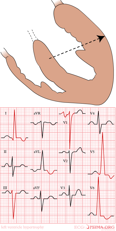 Mechanism of left ventricular hypertrophy

-
-
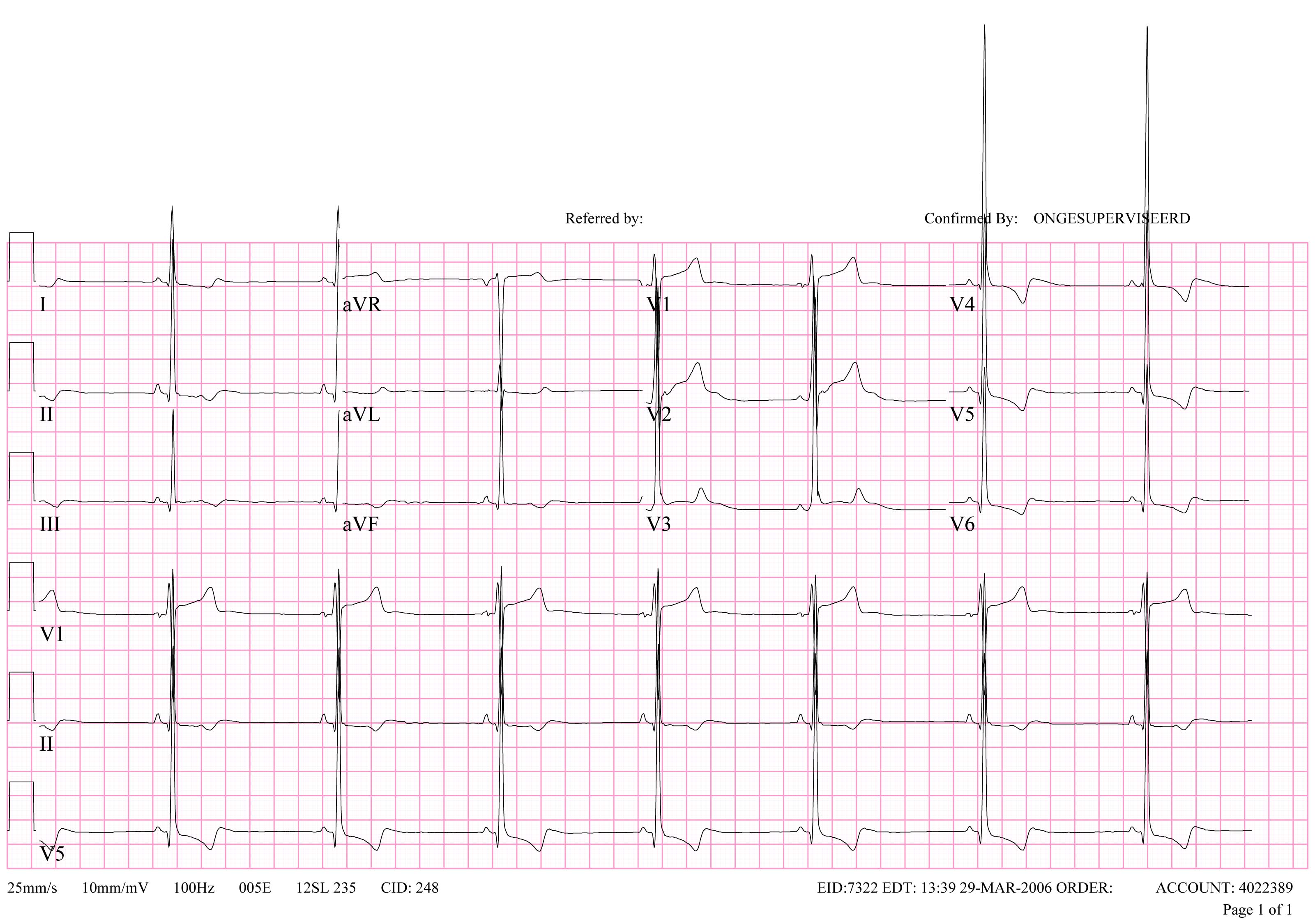
Extreme left ventricular hypertrophy in a patient with severe aortic valve stenosis

-
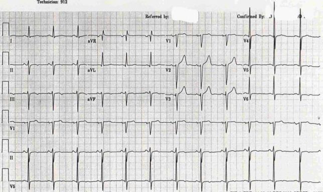
LVH in subendocardial ischemia with positive cardiovascular markers
-
 LVH + Left Anterior Hemiblock

References
- ↑ Sokolow, M, and Lyon, T.P.: The Ventricular Complex As Obtained By Unipolar Limb Leads. Am. Heart J. 1949:37,161.
- ↑ Hammill S. C. Electrocardiographic diagnoses: Criteria and definitions of abnormalities, Chapter 18, MAYO Clinic, Concise Textbook of Cardiology, 3rd edition, 2007 ISBN 0-8493-9057-5
- ↑ Casale, P., Electrocardiographic detection of left ventricular hypertrophy: Development and prospective evaluation of improved criteria. J. Am. Coll Cardiol. 1985:6,572
- ↑ Roberts, W. and Podalak, M: The king of hearts: Analysis of 23 patients with hearts weighing 1,000 grams or more. Am J. Cardiol. 1985:55,485.
- ↑ Surawicz, B.: Electrocardiographic diagnosis of chamber enlargement. J. Am. Coll. Cardiol. 1986: 8,711.
© 2026 MyEClinic – IFTM Institut für Telematik in der Medizin GmbH