
Loop of Henle
Overview
In the kidney, the loop of henle is the portion of the nephron that leads from the proximal convoluted tubule to the distal convoluted tubule. The loop has a hairpin bend in the renal medulla. The main function of this structure is to reabsorb water and ions from the urine. To do this, it uses a countercurrent multiplier mechanism in the medulla. It is named after its discoverer, F. G. J. Henle.
Components
It can be divided into four parts:
| descending limb of loop of Henle | The descending limb has low permeability to ions and urea, while being highly permeable to water. |
| thin ascending limb of loop of Henle | The thin ascending limb is not permeable to water, but it is permeable to ions. |
| medullary thick ascending limb of loop of Henle | Sodium (Na+), potassium (K+) and chloride (Cl–) ions are reabsorbed from the urine by active transport. K+ is passively transported along its concentration gradient through a K+ channel in the basolateral aspect of the cells, back into the lumen of the ascending limb. This K+ “leak” generates a positive electrochemical potential difference in the lumen. The electrical gradient drives more reabsorption of Na+, as well as other cations such as magnesium (Mg2+) and importantly calcium Ca2+. |
| cortical thick ascending limb | The cortical thick ascending limb drains urine into the distal convoluted tubule. |
Blood supply
The loop of Henle is supplied by blood in a series of straight capillaries descending from the cortical efferent arterioles. These capillaries (called the vasa recta; recta is from the Latin for “straight”) also have a countercurrent exchange mechanism that prevents washout of solutes from the medulla, thereby maintaining the medullary concentration. As water is osmotically driven from the descending limb into the interstitium, it readily enters the vasa recta. The low bloodflow through the vasa recta allows time for osmotic equilibration, and can be altered by changing the resistance of the vessels’ efferent arterioles.
Also, the vasa recta still has the large proteins and ions which were not filtered through the glomerulus, which provides an oncotic pressure for ions to enter the vasa recta from the interstitium.
Additional images
-
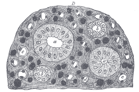 Transverse section of pyramidal substance of kidney of pig, the bloodvessels of which are injected.

References
- Douglas C. Eaton, John Pooler (2004). Vander’s Renal Physiology (6th edition ed.). McGraw-Hill Medical. ISBN 0-07-135728-9.
External links
© 2026 MyEClinic – IFTM Institut für Telematik in der Medizin GmbH