
Myxoma chest x ray
Editor-In-Chief: C. Michael Gibson, M.S., M.D. [1]; Associate Editor-In-Chief: Maria Fernanda Villarreal, M.D. [2] Cafer Zorkun, M.D., Ph.D. [3] Ahmad Al Maradni, M.D. [4]
Overview
There are no specific chest x-ray findings associated with cardiac myxoma, the results can be reported as normal.
Key Chest X-Ray Findings in Cardiac Myxoma
- There are no specific chest x-ray findings associated with cardiac myxoma, the results can be reported as normal.[1]
- Related imaging findings include cardiomegaly, left atrial enlargement, vascular redistribution, prominent pulmonary trunk, and intracardiac tumoral calcification (rare).[2]
Gallery
-
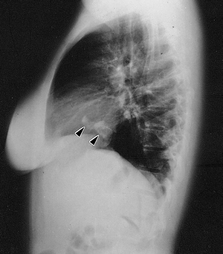 Lateral chest radiograph from a 16-year-old girl with syncope and bacterial endocarditis. The radiograph demonstrates two areas of dense calcification (arrowheads) overlying the posterior aspect of heart. The posterior-anterior (PA) view confirmed location in the heart (not shown). At surgery a calcified myxoma of the right atrium was removed. Image courtesy of Professor Peter Anderson DVM PhD and published with permission © PEIR, University of Alabama at Birmingham, Department of Pathology

| Imaging Technique | Features | Description | Advantages | Limitations |
|---|---|---|---|---|
| Two- or three-dimensional echocardiography |
|
|
|
|
| MRI |
|
|
|
|
| CT |
|
|
|
|
| Angiography |
|
|
|
|
| Chest x-ray |
|
|
|
|
References
- ↑ Cardiac Myxoma. Radiopedia.http://radiopaedia.org/articles/cardiac-myxoma Accessed on November 24, 2015
- ↑ Thyagarajan B, Kumar MP, Patel S, Agrawal A (January 2017). “Extracardiac manifestations of atrial myxomas”. J Saudi Heart Assoc. 29 (1): 37–43. doi:10.1016/j.jsha.2016.07.003. PMC 5247297.
- ↑ Reeder GS, Khandheria BK, Seward JB, Tajik AJ (1991). “Transesophageal echocardiography and cardiac masses”. Mayo Clin. Proc. 66 (11): 1101–9. PMID 1943240.
© 2026 MyEClinic – IFTM Institut für Telematik in der Medizin GmbH