
Nasopharyngeal carcinoma pathophysiology
Editor-In-Chief: C. Michael Gibson, M.S., M.D. [1] Associate Editor(s)-in-Chief: Homa Najafi, M.D.[2]Faizan Sheraz, M.D. [3]
Overview
On microscopic histopathological analysis, abundant dense eosinophilic cytoplasm and prominent lymphoid component are characteristic findings of nasopharyngeal carcinoma.
Pathophysiology
Genetics
Genes involved in the pathogenesis of nasopharyngeal carcinoma include:
Pathology
Gross Pathology
- Nasal cavity involvement – common in early disease[1]
The tumor arises from epithelial cells on the surface of the nasopharynx.
Microscopic Pathology
Features:[2]
- Prominent lymphoid component (Lymphoepithelioma) – key feature
- Features of squamous cell carcinoma:

Nasopharyngeal carcinoma may be classified according to microscopic features into 3 subtypes:[3]
- Well-differentiated (keratinizing type)
- Moderately-differentiated (nonkeratinizing type)
- Undifferentiated (most strongly associated with Epstein-Barr virus infection)
-
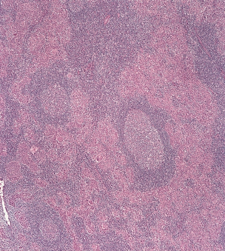 Undifferentiated nasopharyngeal carcinoma – low power
 -
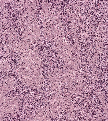 Undifferentiated nasopharyngeal carcinoma – med. power
 -
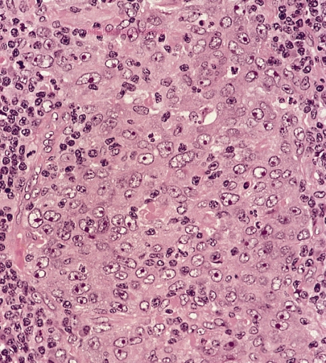 Undifferentiated nasopharyngeal carcinoma – high power


Immunohistochemistry
Immunohistochemistry stains for nasopharyngeal carcinoma include:
- EBER positive
- p16 negative[4]
References
- ↑ Abdel Khalek Abdel Razek, A.; King, A. (2012). “MRI and CT of nasopharyngeal carcinoma”. AJR Am J Roentgenol. 198 (1): 11–8. doi:10.2214/AJR.11.6954. PMID 22194474. Unknown parameter
|month=ignored (help) - ↑ Nasopharyngeal carcinoma http://librepathology.org/wiki/index.php/Nasopharyngeal_carcinoma
- ↑ Richard Cote, Saul Suster, Lawrence Weiss, Noel Weidner (Editor). Modern Surgical Pathology (2 Volume Set). London: W B Saunders. ISBN 0-7216-7253-1.
- ↑ Gulley ML, Nicholls JM, Schneider BG, Amin MB, Ro JY, Geradts J (1998). “Nasopharyngeal carcinomas frequently lack the p16/MTS1 tumor suppressor protein but consistently express the retinoblastoma gene product”. Am. J. Pathol. 152 (4): 865–9. PMC 1858242. PMID 9546345. Unknown parameter
|month=ignored (help)
© 2026 MyEClinic – IFTM Institut für Telematik in der Medizin GmbH