
Papillary muscle
Editor-In-Chief: C. Michael Gibson, M.S., M.D. [1];Associate Editor-In-Chief: Cafer Zorkun, M.D., Ph.D. [2]
Overview
In anatomy, the papillary muscles of the heart serve to limit the movements of the mitral and tricuspid valves. These muscles contract to tighten the chordae tendineae, which in turn prevent inversion. This occurs in response to pressure gradients. Instead they brace the valves against the high pressure, preventing regurgitation of ventricular blood back into the atrial cavities.
-
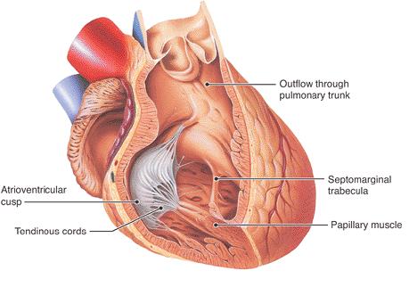 Image courtesy of Hartwig, Walter C., The Editor of Fundamental Anatomy, 1st Edition, 2008 and copyrighted
 -
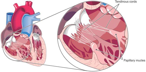 Image courtesy of Hartwig, Walter C., The Editor of Fundamental Anatomy, 1st Edition, 2008 and copyrighted


Additional images
-
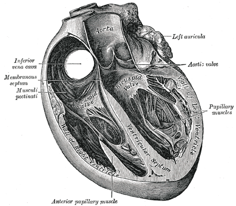 Section of the heart showing the ventricular septum.
 -
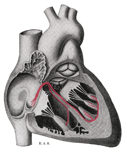 Schematic representation of the atrioventricular bundle of His.


References
© 2026 MyEClinic – IFTM Institut für Telematik in der Medizin GmbH