
Pyelonephritis CT scan
Editor-In-Chief: C. Michael Gibson, M.S., M.D. [1]; Associate Editor(s)-in-Chief: Usama Talib, BSc, MD [2]
Overview
A CT scan can be used to detect diffuse or complicated pyelonephritis and its suspected complications. It is used when the suspicion of pyelonephritis is accompanied by other differentials. CT scan is very sensitive and CT urography is sometimes used for imaging of the urinary tract. The extent of damage to the parenchymal tissue can also be witnessed in detail with a CT scan.
CT
Findings on a CT scan may vary with respect to the type of pyelonephritis. A CT scan usually demonstrates:[1][2][3][4][5][6]
Acute Pyelonephritis
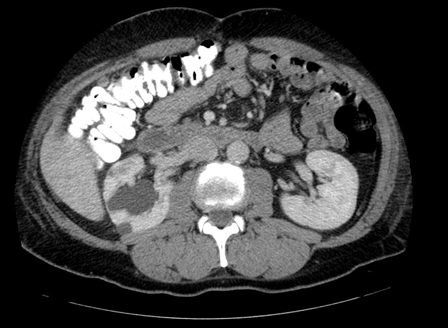
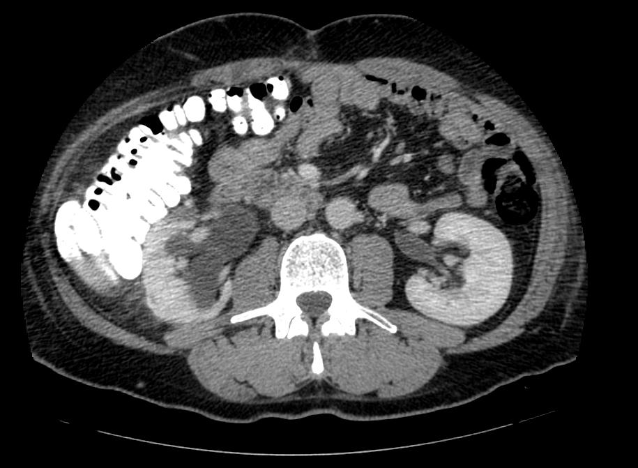
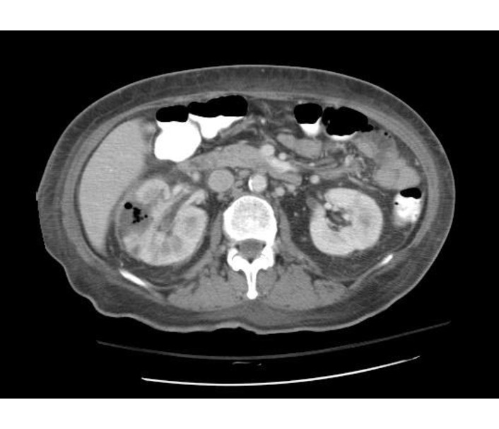
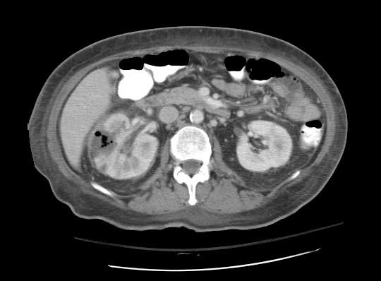
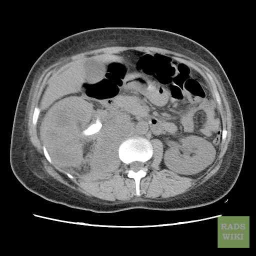
CT scan in case of acute pyelonephritis may demonstrate:[1][7] Images courtesy of RadsWiki
-
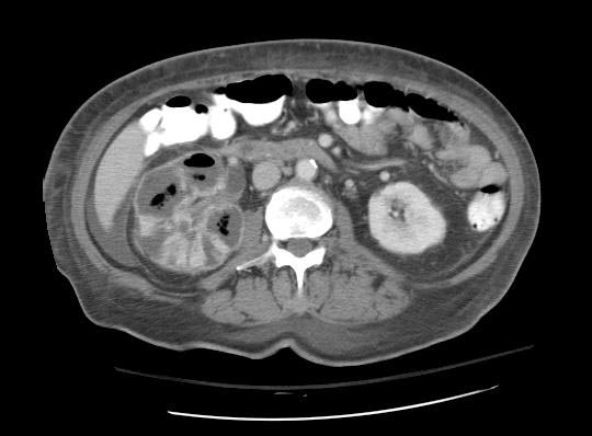
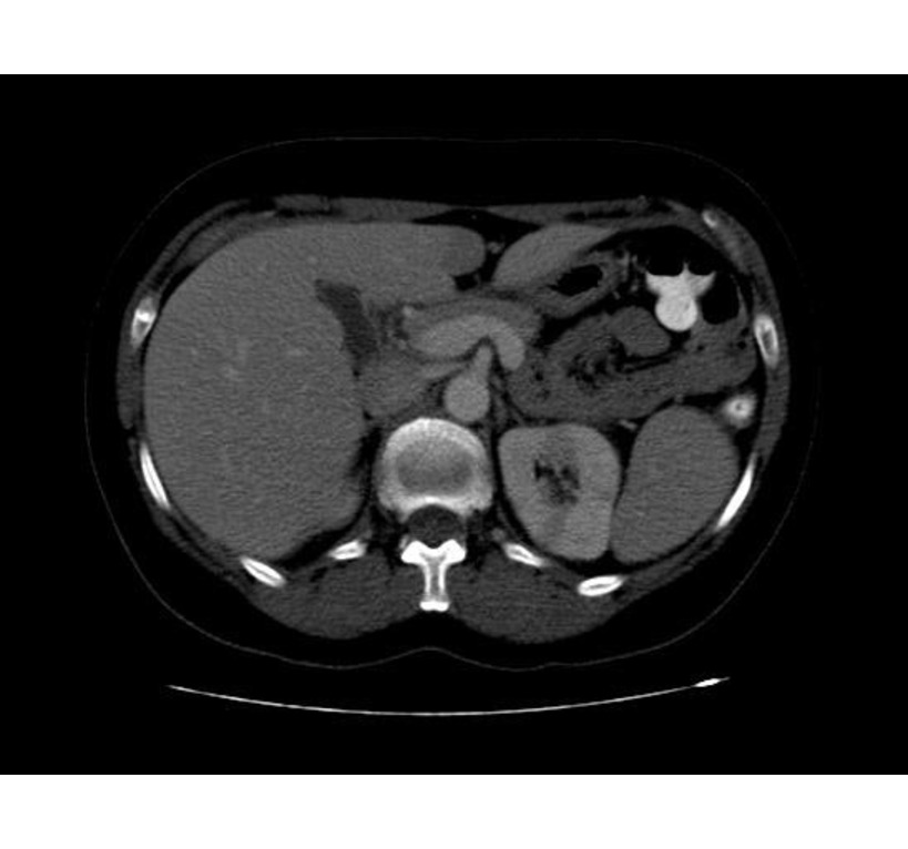 CT: Acute pyelonephritis
 -
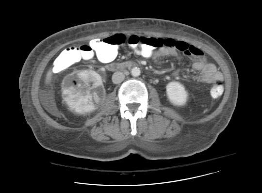
 CT: Acute pyelonephritis
 -
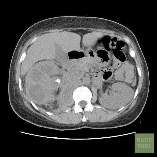
 CT: Acute pyelonephritis


Chronic Pyelonephritis
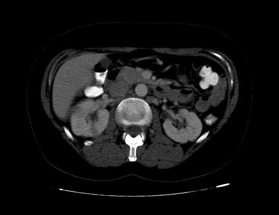
Imaging findings are characterized by renal scarring, atrophy and cortical thinning, hypertrophy of residual normal tissue, caliceal clubbing secondary to retraction of the papilla from overlying scar, thickening and dilatation of the caliceal system, and overall renal asymmetry. Images courtesy of RadsWiki
-
 CT image demonstrates chronic pyelonephritis on the right
 -
 CT image demonstrates chronic pyelonephritis on the right
 -
 CT image demonstrates chronic pyelonephritis on the right


Emphysematous Pyelonephritis
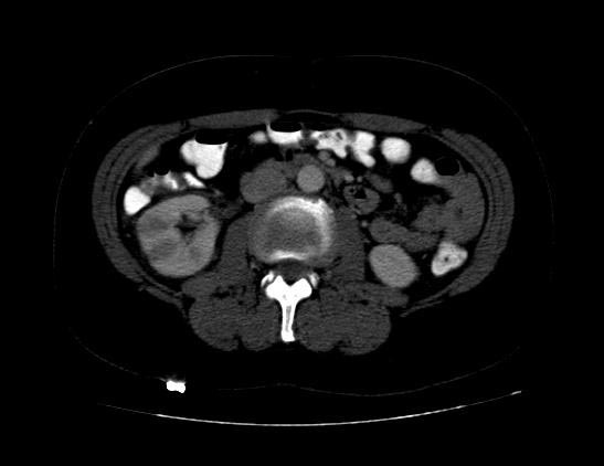
- Additional evaluation with CT will confirm the presence and extent of parenchymal gas and will often allow identification of the source of obstruction when present.
- The use of intravenous contrast material will often reveal asymmetric renal enhancement or delayed excretion, and, during the nephrographic phase, will help identify areas of focal tissue necrosis or abscess formation.
-
 CT: Emphysematous pyelonephritis
 -
 CT: Emphysematous pyelonephritis
 -
 CT: Emphysematous pyelonephritis
 -
 CT: Emphysematous pyelonephritis


Xanthogranulomatous Pyelonephritis
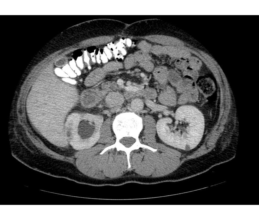
The CT findings of xanthogranulomatous pyelonephritis are pathognomonic in most cases: diffuse reniform enlargement with ill-defined central low attenuation, apparent cortical thinning, and central calculi.[8]
- Extension into the perinephric space and beyond the Gerota fascia is not uncommon.
- Central areas of low attenuation represent nonenhancing xanthomatous material that may demonstrate attenuation values less than those of water.
-
 CT image demonstrates right xanthogranulomatous pyelonephritis
 -
 CT image demonstrates right xanthogranulomatous pyelonephritis
 -
 CT image demonstrates right xanthogranulomatous pyelonephritis
 -
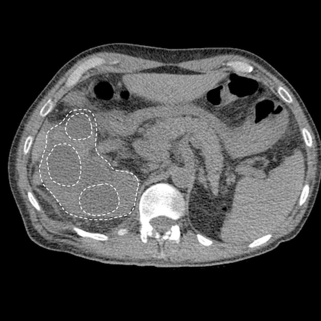 CT image demonstrates right xanthogranuomatous pyelonephritis with dilated calyces


References
- ↑ 1.0 1.1 Meyrier A, Condamin MC, Fernet M, Labigne-Roussel A, Simon P, Callard P; et al. (1989). “Frequency of development of early cortical scarring in acute primary pyelonephritis”. Kidney Int. 35 (2): 696–703. PMID 2651759.
- ↑ Soulen MC, Fishman EK, Goldman SM (1989). “Sequelae of acute renal infections: CT evaluation”. Radiology. 173 (2): 423–6. doi:10.1148/radiology.173.2.2798873. PMID 2798873.
- ↑ Demertzis J, Menias CO (2007). “State of the art: imaging of renal infections”. Emerg Radiol. 14 (1): 13–22. doi:10.1007/s10140-007-0591-3. PMID 17318482.
- ↑ Fowler JE, Perkins T (1994). “Presentation, diagnosis and treatment of renal abscesses: 1972-1988”. J Urol. 151 (4): 847–51. PMID 8126807.
- ↑ Gupta K, Hooton TM, Naber KG, Wullt B, Colgan R, Miller LG; et al. (2011). “International clinical practice guidelines for the treatment of acute uncomplicated cystitis and pyelonephritis in women: A 2010 update by the Infectious Diseases Society of America and the European Society for Microbiology and Infectious Diseases”. Clin Infect Dis. 52 (5): e103–20. doi:10.1093/cid/ciq257. PMID 21292654.
- ↑ Kawashima A, LeRoy AJ (2003). “Radiologic evaluation of patients with renal infections”. Infect Dis Clin North Am. 17 (2): 433–56. PMID 12848478.
- ↑ Tsugaya M, Hirao N, Sakagami H, Iwase Y, Watase H, Ohtaguro K; et al. (1990). “Computerized tomography in acute pyelonephritis: the clinical correlations”. J Urol. 144 (3): 611–3. PMID 2388315.
- ↑ Radiopaedia.org. Case courtesy of A.Prof Frank Gaillard, <a href=”https://radiopaedia.org/“>Radiopaedia.org</a>. From the case <a href=”https://radiopaedia.org/cases/9931“>rID: 9931
© 2026 MyEClinic – IFTM Institut für Telematik in der Medizin GmbH