
Renal vein
Editor-In-Chief: C. Michael Gibson, M.S., M.D. [1]
The renal veins are veins that drain the kidney. They connect the kidney to the inferior vena cava.
It is usually singular to each kidney, except in the condition “multiple renal veins”.[1]
Asymmetry
Because the inferior vena cava is on the right half of the body, the left renal vein is generally the longer of the two.
Because the inferior vena cava is not laterally symmetrical, the left renal vein often receives the following veins:[2]
- left inferior phrenic vein
- left suprarenal vein
- left gonadal vein (left testicular vein in males, left ovarian vein in females)
- left 2nd lumbar vein
This is in contrast to the right side of the body, where these veins drain directly into the IVC.
Pathology
Diseases associated with the renal vein include renal vein thrombosis (RVT) and nutcracker syndrome (renal vein entrapment syndrome).
References
Additional images
-
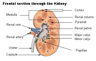 Frontal section through the kidney
 -
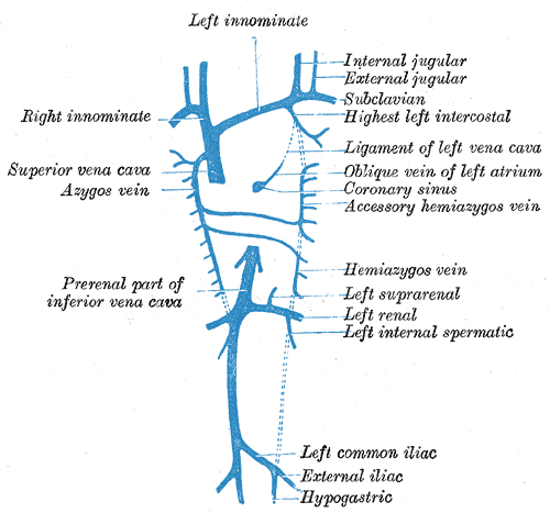 Diagram showing completion of development of the parietal veins.
 -
 The venæ cavæ and azygos veins, with their tributaries.


-
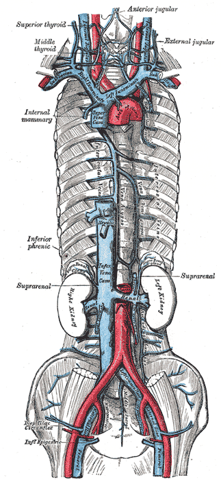
 Human kidneys viewed from behind with spine removed.

See also
Reference
External links
- Template:SUNYAnatomyFigs – “Retroperitoneal structures on the posterior abdominal wall.”
© 2026 MyEClinic – IFTM Institut für Telematik in der Medizin GmbH