
Superior vena cava syndrome CT
Editor-In-Chief: C. Michael Gibson, M.S., M.D. [1]Associate Editor(s)-in-Chief: Maria Fernanda Villarreal, M.D. [2]
Overview
On enhanced CT scan, findings include location and severity of the superior vena cava obstruction, superimposed thrombosis, a mediastinal mass or lymphadenopathy, collateral vessels, and associated lung masses. CT scan is the imaging modality of choice.
CT
- CT scan is the imaging modality of choice for the diagnosis of superior vena cava syndrome.
- On CT scan, superior vena cava syndrome is characterized by:[1]
- Location and severity of the superior vena cava obstruction
- Superimposed thrombosis
- Mediastinal mass or lymphadenopathy
- Collateral vessels and associated lung masses
Gallery
-
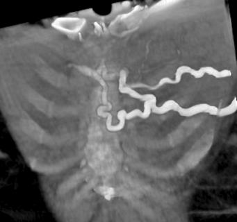 Collaterals on CT scan in a patient with superior vena cava syndrome

-
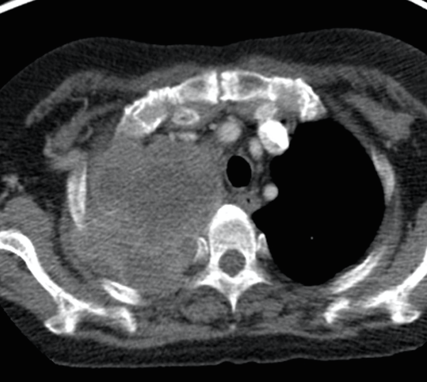 Lung cancer with vena cava invasion.

References
- ↑ Superior Vena Cava Syndrome.Dr Amir Rezaee and Radswiki et al. Radiopedia http://radiopaedia.org/articles/superior-vena-cava-obstruction Accessed on January 13, 2016
© 2026 MyEClinic – IFTM Institut für Telematik in der Medizin GmbH