
Thromboembolism other imaging findings
Editor-In-Chief: C. Michael Gibson, M.S., M.D. [1]
Other Imaging Findings
Contrast Venography
Contrast venography (also called Venography or phlebography) is the definitive test for diagnosing deep venous thrombosis which taken after a special dye is injected into the vein or even bone marrow.
Contrast venography can also help;
- to distinguish blood clots from obstructions in the veins
- to evaluate congenital vein problems
- to evaluate veins prior to treatment of chronic venous insufficiency
- to control functioning of deep leg vein valves
- to identify a vein graft for coronary artery bypass surgery
-
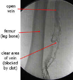 Venography: Deep venous thrombosis. Source
 -
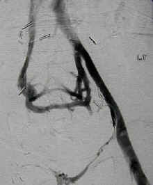 An occluded vein with collateral vessel formation. Source


Pulmonary Angiography
Pulmonary angiography (or pulmonary arteriography) is a cardiological medical procedure. Pulmonary arteries are visualized to detect blood clots (such as a pulmonary embolism) or arteriovenous malformations.
The use of pulmonary angiography has been largely replaced by spiral CT in diagnosis of pulmonary embolism.
-
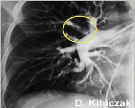 Pulmonary angiogram in a patient with pulmonary embolus. A thrombus is observed in the area within the yellow circle. Source

Ventilation / Perfusion Scan
Ventilation/perfusion scan (or V/Q scan or lung scintigraphy), which shows that some areas of the lung are being ventilated but not perfused with blood (due to obstruction by a clot). This type of examination is used less often because of the more widespread availability of CT technology, however, it may be useful in patients who have an allergy to iodinated contrast or in pregnancy due to lower radiation exposure than CT. * The ventilation/perfusion ratio (V/Q) Scan: The PIOPED data suggests that normal perfusion scans are almost never associated with recurrent pulmonary embolism, even if anticoagulation is withheld.
References
© 2026 MyEClinic – IFTM Institut für Telematik in der Medizin GmbH