
Alveolar duct
Editor-In-Chief: C. Michael Gibson, M.S., M.D. [1]
Overview
Overview
Alveolar ducts are the tiny end ducts of the branching airways that fill the lungs. Each lung holds approximately 1.5 to 2 million of them. The tubules divide into two or three alveolar sacs at the distal end. They are formed from the confluence openings of several alveoli. Distal terminations of alveolar ducts are atria which then end in alveolar sacs.
In human anatomy, respiratory bronchioles exists proximal to the alveolar ducts. The epithelial lining consists of smooth muscle knobs covered by nonciliated, simple cuboidal cells. The smooth muscle constricts under parasympathetic innervation and relax under sympathetic innervation.
Other Imaging Findings
Other Imaging Findings
-
![Detailed drawing of the alveoli from Gray's Anatomy, 1918 - Schematic longitudinal section of a primary lobule of the lung (anatomical unit); r. b respiratory bronchiole; al. d alveolar duct; at atria; a. s alveolar sac; 'a' alveolus or air cell; p. a.: pulmonary artery; p. v pulmonary vein; l lymphatic; l. n lymph node.]](https://www.wikidoc.org/images/8/8f/Gray975.png) Detailed drawing of the alveoli from Gray’s Anatomy, 1918 – Schematic longitudinal section of a primary lobule of the lung (anatomical unit);
Detailed drawing of the alveoli from Gray’s Anatomy, 1918 – Schematic longitudinal section of a primary lobule of the lung (anatomical unit);
r. b respiratory bronchiole;
al. d alveolar duct; at atria;
a. s alveolar sac;
‘a’ alveolus or air cell;
p. a.: pulmonary artery;
p. v pulmonary vein;
l lymphatic;
l. n lymph node.] -
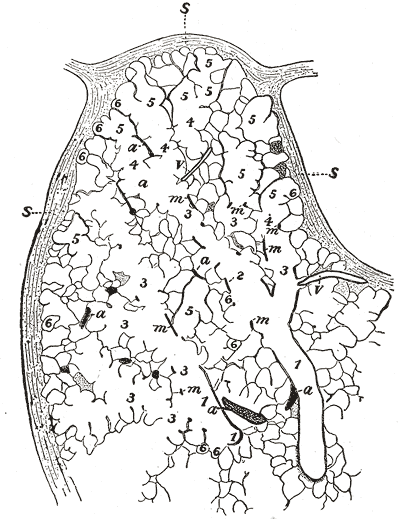 Part of a secondary lobule from the depth of a human lung, showing parts of several primary lobules.
1, bronchiole;
2, respiratory bronchiole;
3, alveolar duct;
4, atria;
5, alveolar sac;
6, alveolus or air cell:
m, smooth muscle;
a, branch pulmonary artery;
v, branch pulmonary vein;
s, septum between secondary lobules. -
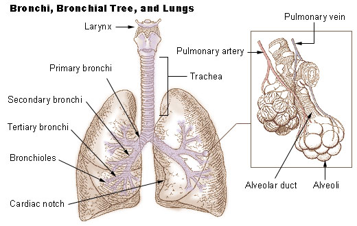 Bronchi, bronchial tree, and lungs
 -
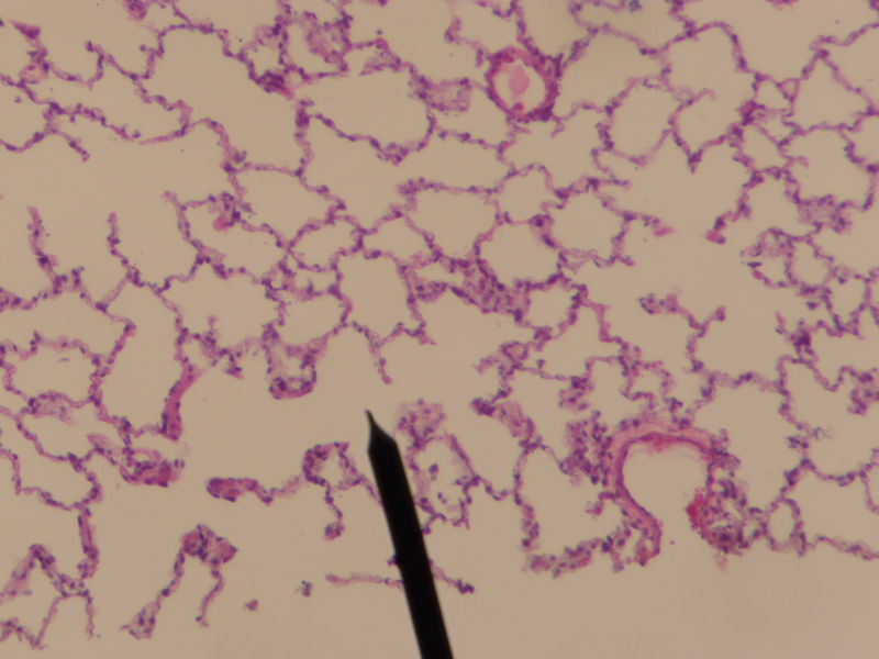 Human alveolar duct
![Detailed drawing of the alveoli from Gray's Anatomy, 1918 - Schematic longitudinal section of a primary lobule of the lung (anatomical unit); r. b respiratory bronchiole; al. d alveolar duct; at atria; a. s alveolar sac; 'a' alveolus or air cell; p. a.: pulmonary artery; p. v pulmonary vein; l lymphatic; l. n lymph node.]](https://www.wikidoc.org/index.php/File%3AGray975.png)


Looking for the patient version?
© 2026 MyEClinic – IFTM Institut für Telematik in der Medizin GmbH