
Angiomyolipoma other imaging findings
Editor-In-Chief: C. Michael Gibson, M.S., M.D. [1] Associate Editor(s)-in-Chief: Faizan Sheraz, M.D. [2],Rekha, M.D.
Overview
Overview
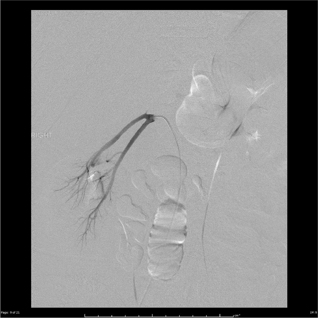
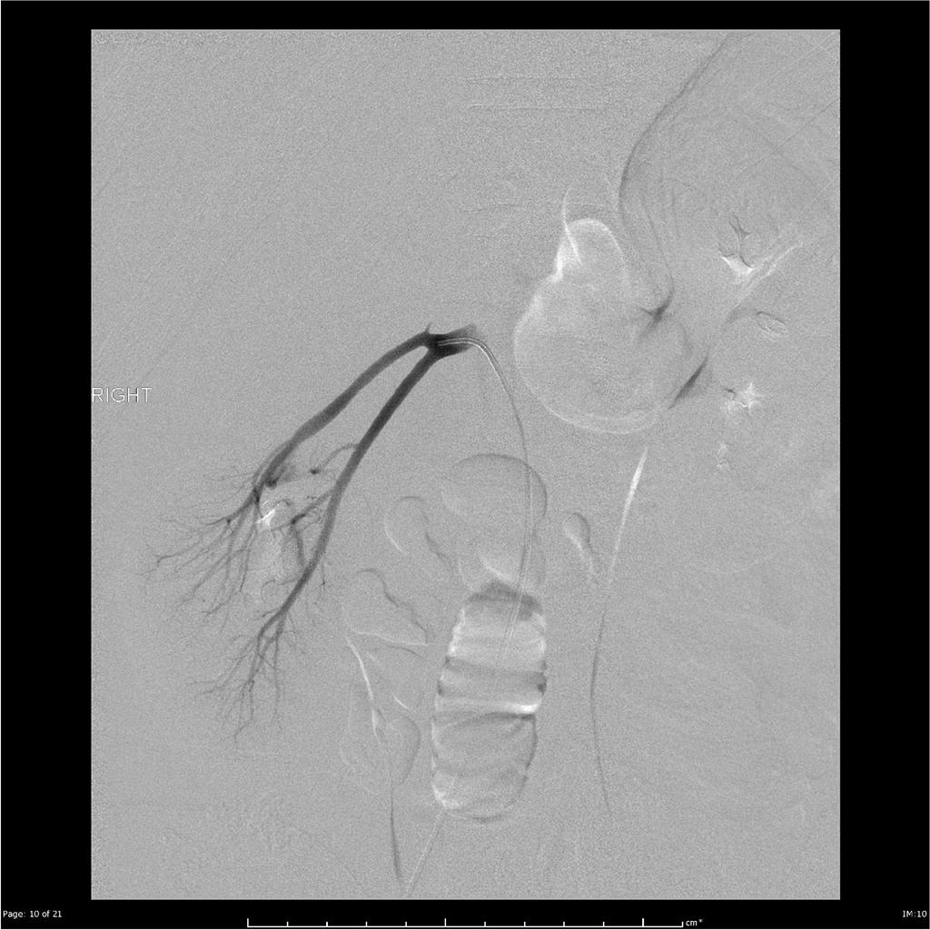
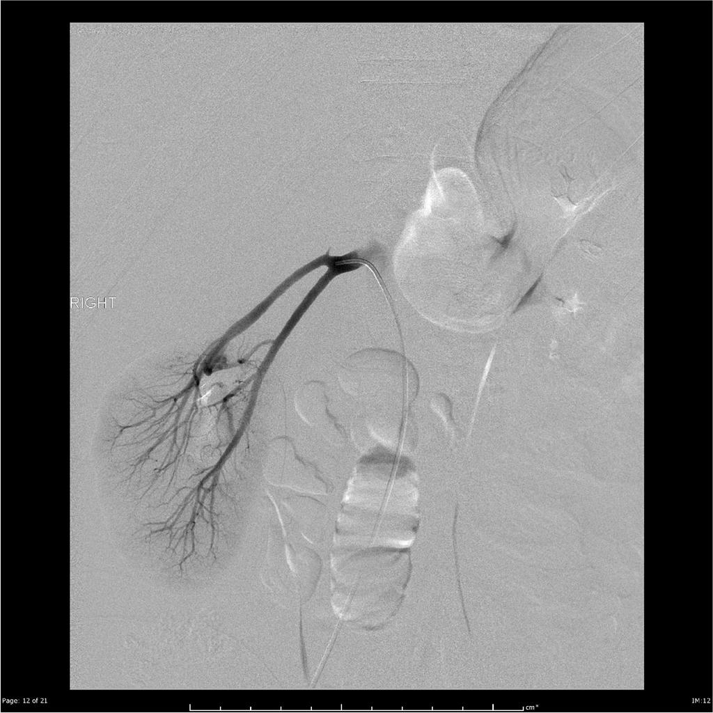
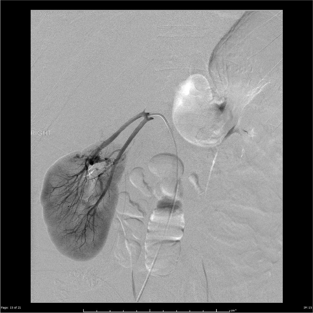
On digital subtraction angiography, angiomyolipoma is characterized by hypervascular lesions, microvascular aneurysms, and absent AV shunting.
Other Imaging Findings
Other Imaging Findings
Digital Subtraction Angiography
On digital subtraction angiography, angiomyolipoma is characterized by:
- Hypervascular lesions
- Microaneurysms or macroaneurysms
- Sharply marginated
- Dense early arterial network
- Late whorled appearance
- Absent AV shunting
-
 A
A -
 B
B -
 C
C -
 D
D


- Figures A-D: Angiographic runs showed a significantly ptosed right kidney, with supply to the large upper pole angiomyolipoma via tortuous artery arising from the superior division of the right renal artery.[1]
References
References
- ↑ Angiomyolipoma Image courtesy of Dr. Andrew LawsonRadiopaedia(original file “here”). Creative Commons BY-SA-NC
Looking for the patient version?
© 2026 MyEClinic – IFTM Institut für Telematik in der Medizin GmbH