
Aortic arch anomalies CT
Editor-In-Chief: C. Michael Gibson, M.S., M.D. [1]
Associate Editor-In-Chief: Cafer Zorkun, M.D., Ph.D. [2] Keri Shafer, M.D. [3] Priyamvada Singh, MBBS [[4]]
Assistant Editor-In-Chief: Kristin Feeney, B.S. [[5]]
Overview
Overview
CT
CT
Computed tomography can be used as a diagnostic tool to show the relationship of the aortic arches to the trachea and esophagus and also the degree of tracheal narrowing. Bronchoscopy can be useful in internally assessing the degree of tracheomalacia.
Imaging Findings
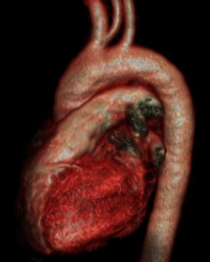
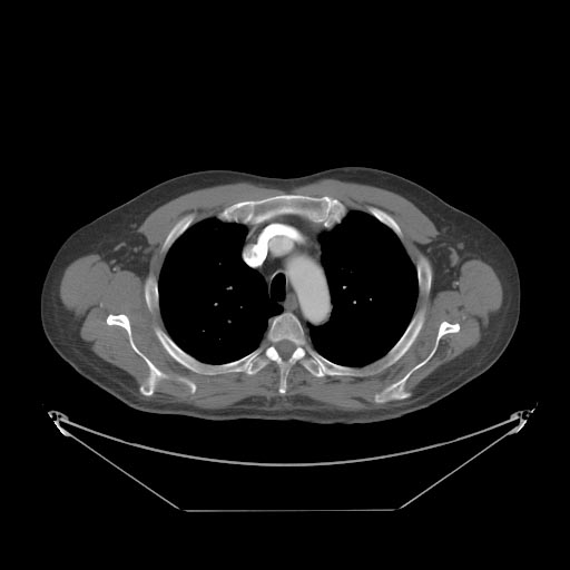
Classification: Double aortic arch
Case: A chest CT was performed in the arterial phase. A diagram of the findings is seen. The left common carotid artery arose from the ascending aorta. The aortic arch was to the right of the trachea, and passed posterior to it. The right common carotid and right subclavian arteries arose from the arch in that order. From the posterior arch, the left subclavian artery arose from a large diverticulum (this corresponds to the left “aortic knuckle” seen on the PA film). The descending aorta then crossed to the right again before passing through the diaphragm in the midline. This appearance may be due to right aortic arch with aberrant left subclavian artery, or to double aortic arch with atresia of the left arch, subtype 3. This case is more likely to be the former, as there was no narrowing of the trachea, and subtype 3 double aortic arch is rare. The anatomic difference between these two anomalies is persistence or not of an atretic segment of the left arch.

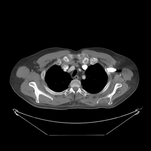
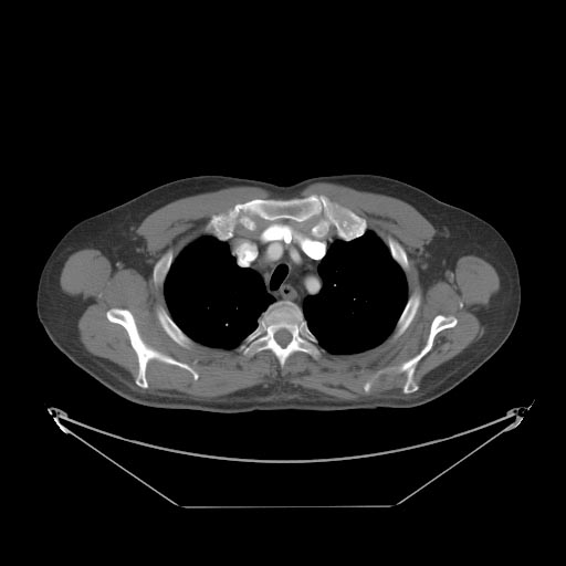
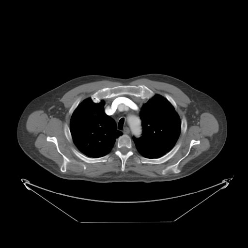
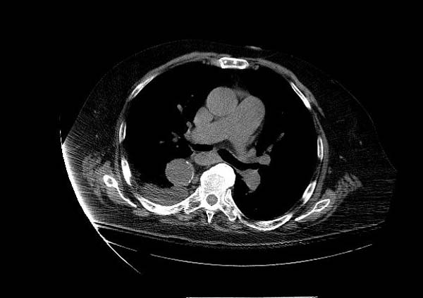
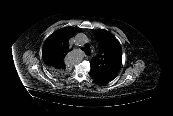
Classification: Bovine arch
Case: A multi-slice CT was performed to deteremine the relationship of the aortic arches.
-
 Bovine arch
 -
 MSCT: Bovine arch
 -
 MSCT: Bovine arch
 -
 MSCT: Bovine arch
 -
 MSCT: Bovine arch
 -
 MSCT: Bovine arch
 -
 Right Aortic Arch
 -
 Right Aortic Arch


Looking for the patient version?
© 2026 MyEClinic – IFTM Institut für Telematik in der Medizin GmbH