
Arachnoid cyst MRI
Editor-In-Chief: C. Michael Gibson, M.S., M.D. [1] Associate Editor(s)-in-Chief: José Eduardo Riceto Loyola Junior, M.D.[2]
Overview
Overview
On brain/spine MRI, arachnoid cysts are characterized by cystic images with similar density to CSF and non-enhancing borders, mostly found in the middle cranial fossa while they only rarely occur in the spinal cord. MRIs are more adequate than CT scans for evaluating arachnoid cysts.
MRI
MRI
- MRIs are better to diagnose and evaluate the extent of the arachnoid cyst than the CT Scan;
- Most cysts (50-60%) are found in the floor of the middle cranial fossa, while 1/4 to 1/3 of occur in the posterior fossa, particularly in the retrocerebellar, cerebellopontine, and quadrigeminal plate cisterns. Rarely, they may be found in the spinal cord.[1][2]
- Demonstrate the exact location, extent, and relationship of the cyst;
- Can differentiate arachnoid from epidermoid cysts (arachnoid cysts are identical to CSF, while epidermoid present a higher signal with FLAIR and reduced diffusion with DWI, making them appear brighter than CSF).
- CSF signal is seen within the cyst;
- Eventually, arachnoid cysts may contain proteinaceous fluid or blood, which can cause diagnostic confusion.[3]
Differential Diagnosis
| Intraventricularly: | Colloid cysts |
| Intraparenchymally: | Parasitic infections, cystic metastases |
| Porencephalic cysts | |
| Craniopharyngiomas | |
| Holoprosencephalies | |
| Agenesis of corpus callosum | |
| Defect in the hemispheral cleavage | |
| Dandy-Walker complex (posterior fossa cysts) |
As differential diagnosis, the following hypothesis must be considered:
- Enlarged CSF space;
- Epidermoid cyst;
- Subdural hygroma/chronic subdural hemorrhage;
- Cystic tumors;
- Pilocytic astrocytoma;
- Hemangioblastoma;
- Neurenteric cyst;
- Neuroglial cyst;
- Porencephalic cyst;
- Neurocysticercosis.[5]
References
References
- ↑ Robertson, S. J., S. M. Wolpert, and V. M. Runge. “MR imaging of middle cranial fossa arachnoid cysts: temporal lobe agenesis syndrome revisited.” American journal of neuroradiology 10.5 (1989): 1007-1010.
- ↑ “Arachnoid Cysts – Imaging”. Medscape. 06/26/2020. Check date values in:
|date=(help) - ↑ “Arachnoid Cysts”. MedPix. 06/26/2020. Check date values in:
|date=(help) - ↑ Cincu, Rafael, Amit Agrawal, and Jose Eiras. “Intracranial arachnoid cysts: current concepts and treatment alternatives.” Clinical neurology and neurosurgery 109.10 (2007): 837-843.
- ↑ “Arachnoid Cysts – Radiopaedia”. Radiopaedia. 06/26/2020. Check date values in:
|date=(help)
MRI Examples of Arachnoid Cysts
MRI Examples of Arachnoid Cysts
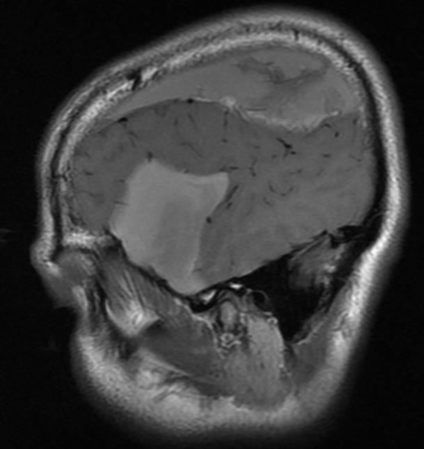
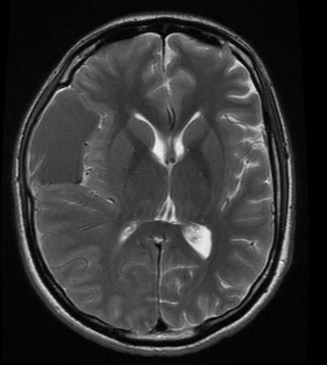
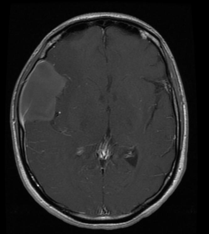
Patient #1: Left middle cranial fossa arachnoid cyst
-
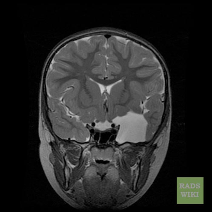 Cor T2
 -
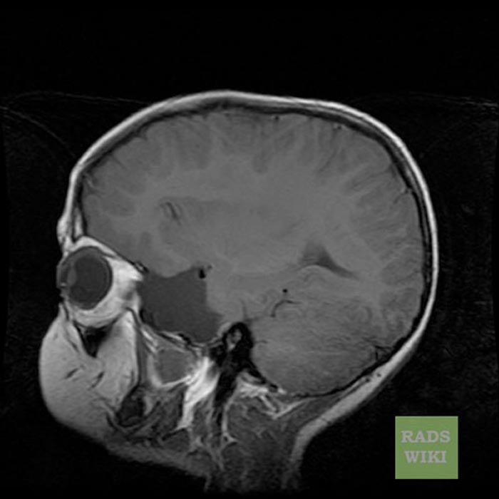 Sag T1
 -
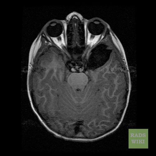 Axial T1 FLAIR
 -
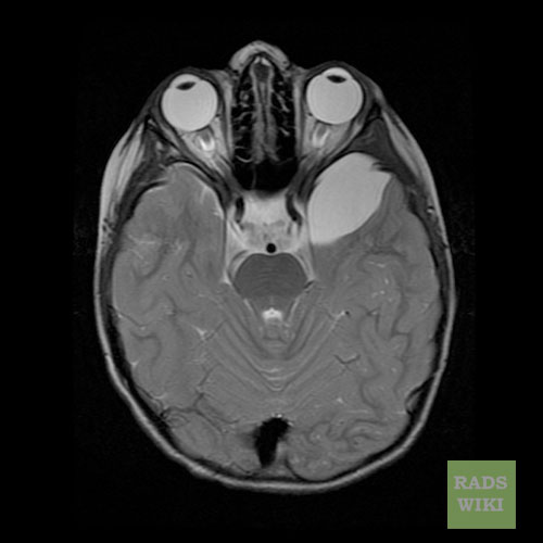 Axial T2


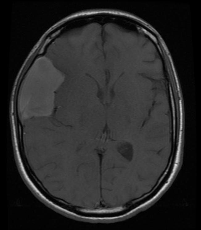
Patient #2: CT and MR images demonstrate a hemorrhagic arachnoid cyst
-
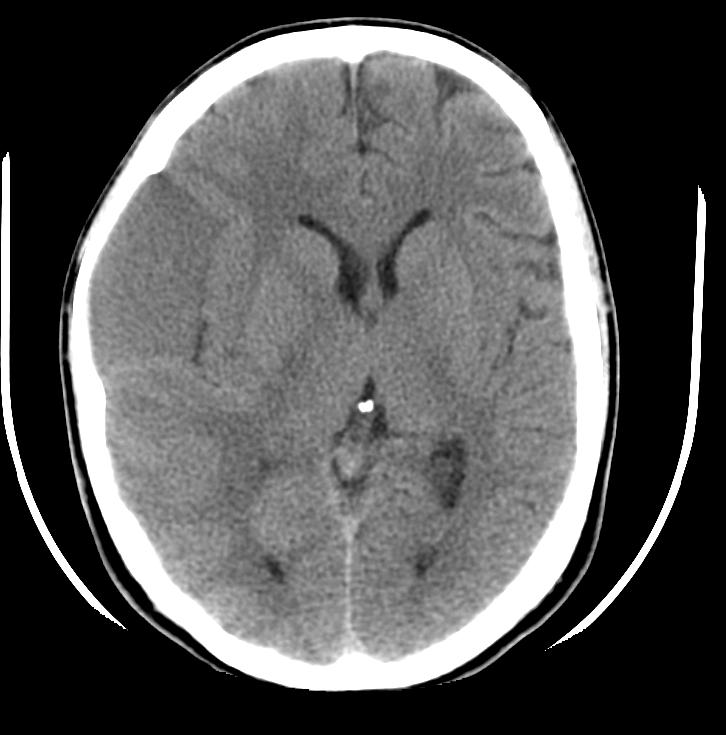 CT
CT -
 Sag T1
 -
 Ax T1
Ax T1 -
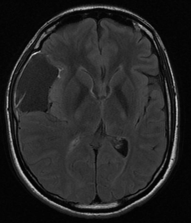 Ax FLAIR
 -
 Ax T2
Ax T2 -
 Ax T1 with GAD


Looking for the patient version?
© 2026 MyEClinic – IFTM Institut für Telematik in der Medizin GmbH