
Biliary cystadenoma and cystadenocarcinoma CT
Editor-In-Chief: C. Michael Gibson, M.S., M.D. [1];Associate Editor(s)-in-Chief: Suveenkrishna Pothuru, M.B,B.S. [2]
Overview
Overview
On CT scan, biliary cystadenoma is characterized by fluid filled multiloculated cyst with calcification of septa and cyst wall. The appearance of the cyst fluid on CT is variable depending on its composition.
CT
CT
Biliary cystadenomas often are diagnosed incidentally, during imaging studies such as ultrasound or CT scan. Both abdominal ultrasound and CT scan are considered the most useful radiologic studies, allowing correct diagnosis in most cases:[1]
- CT scan usually shows a multiloculated cyst.
- Calcification of septa or cyst wall may be observed.
- Calcification that may be present in the septa or cyst wall are typically more apparent with CT than other imaging modalities
- The septa may enhance following administration of contrast.
- As is the case with ultrasound, the appearance of the cyst fluid on CT is variable depending on its composition. It can range from that of water (HU = 0) to quite hyperattenuating if the cyst has been complicated by recent hemorrhage.
There are no specific imaging features that permit reliable differentiation of biliary cystadenoma from cystadenocarcinoma:[2]
- The presence of intraluminal polypoid projections originating from the wall should raise the suspicion for cystadenocarcinoma.[1]
- Hypervascularity of mural nodules on CT also suggests malignancy.[3]
(Images courtesy of RadsWiki)
-
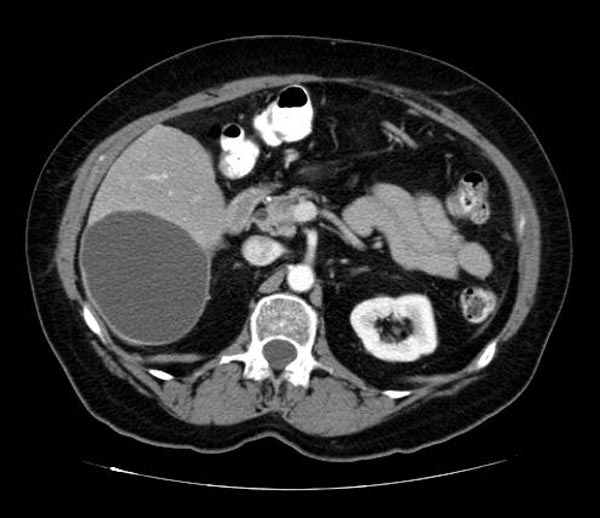 CT image demonstates a biliary cystadenoma
 -
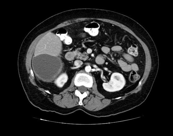 CT image demonstates a biliary cystadenoma


References
References
- ↑ 1.0 1.1 Ramacciato, Giovanni; Nigri, GiuseppeR; D’Angelo, Francesco; Aurello, Paolo; Bellagamba, Riccardo; Colarossi, Cristina; Pilozzi, Emanuela; Del Gaudio, Massimo (2006). World Journal of Surgical Oncology. 4 (1): 76. doi:10.1186/1477-7819-4-76. ISSN 1477-7819. Missing or empty
|title=(help) - ↑ Biliary cystadenoma.Dr Yuranga Weerakkody and Radswiki et al.Radiopaedia.org 2015. http://radiopaedia.org/articles/biliary-cystadenoma
- ↑ Ahanatha Pillai, Sastha; Velayutham, Vimalraj; Perumal, Senthilkumar; Ulagendra Perumal, Srinivasan; Lakshmanan, Anand; Ramaswami, Sukumar; Ramasamy, Ravi; Sathyanesan, Jeswanth; Palaniappan, Ravichandran; Rajagopal, Surendran (2012). “Biliary Cystadenomas: A Case for Complete Resection”. HPB Surgery. 2012: 1–6. doi:10.1155/2012/501705. ISSN 0894-8569.
Looking for the patient version?
© 2026 MyEClinic – IFTM Institut für Telematik in der Medizin GmbH