

Health Dictionary
Reliable, easy-to-read patient information on common medical conditions — written in plain language, free of jargon, and grounded in current evidence. Browse the most frequent diagnoses, understand what they mean for everyday life, and learn which steps and questions help you have a focused conversation with your doctor.
Blepharitis physical examination
Introduction
Overview
Overview
Physical examination of patients with blepharitis is usually remarkable for irritated eyelid edges, hard crusting of the lashes, entropion, ectropion, poliosis, and diffuse conjunctival injection.[1][2]
Physical Examination
Physical Examination
Skin
Common skin examination findings of blepharitis include:[1][2][3]
- Scalp or facial skin itching and flaking (suggestive of seborrheic dermatitis)
- Facial flushing and Telangiectasia on cheeks and nose (suggestive of acne rosacea)
Eye
Gross Eye Examination
Common eye examination findings of blepharitis include:[2][4][5]
- Pink or irritated eyelid edges
- Hard crusting of the lashes (staphylococcal blepharitis)
- Greasy appearing flakes (seborrheic blepharitis)
- Cylindrical dandruff on the base of lashes (suggestive of Demodex folliculorum infestation)
- Telangiectasia on the anterior eyelid
- Entropion
- Ectropion
- Trichiasis
- Madarosis
- Poliosis
- Distichiasis
- Diffuse conjunctival injection
Slit Lamp Examination
On slit lamp examination, blepharitis is characterized by:
- Meibomian gland enlargement
- Waxy secretions at the gland openings
- Lid margin neovascularization and dilation of existing blood vessels
- Ulcerations along the eyelid margin
- Corneal nodules
- Corneal margin ulcer
- Superficial corneal pannus
Images
Images
The following are images associated with blepharitis:[6]
-
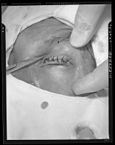 Entropion and trichiasis – By Otis Historical Archives Nat’l Museum of Health & Medicine – originally posted to Flickr as A44-652-11, CC BY 2.0, https://commons.wikimedia.org/w/index.php?curid=6215100

References
References
- ↑ 1.0 1.1 Dougherty JM, McCulley JP (1986). “Bacterial lipases and chronic blepharitis”. Invest Ophthalmol Vis Sci. 27 (4): 486–91. PMID 3957566.
- ↑ 2.0 2.1 2.2 THYGESON P, VAUGHAN DG (1954). “Seborrheic blepharitis”. Trans Am Ophthalmol Soc. 52: 173–88. PMC 1312591. PMID 13274422.
- ↑ Lemp MA, Nichols KK (2009). “Blepharitis in the United States 2009: a survey-based perspective on prevalence and treatment”. Ocul Surf. 7 (2 Suppl): S1–S14. PMID 19383269.
- ↑ Blepharitis. American Academy of Ophthalmology/eyewiki (2014). http://eyewiki.org/Blepharitis Accessed on July 14, 2016
- ↑ Blepharitis. National Eye Institute. (2009). https://nei.nih.gov/health/blepharitis/blepharitis Accessed on July, 14, 2016
- ↑ Tomlinson A, Bron AJ, Korb DR, Amano S, Paugh JR, Pearce EI; et al. (2011). “The international workshop on meibomian gland dysfunction: report of the diagnosis subcommittee”. Invest Ophthalmol Vis Sci. 52 (4): 2006–49. doi:10.1167/iovs.10-6997f. PMC 3072162. PMID 21450918.
Editor-In-Chief: C. Michael Gibson, M.S., M.D. [1]; Associate Editor(s)-in-Chief: Sara Mehrsefat, M.D. [2]