

Health Dictionary
Reliable, easy-to-read patient information on common medical conditions — written in plain language, free of jargon, and grounded in current evidence. Browse the most frequent diagnoses, understand what they mean for everyday life, and learn which steps and questions help you have a focused conversation with your doctor.
Boerhaave syndrome chest x ray
Introduction
Overview
Overview
An upright anterior-posterior view of the chest is the most useful in early diagnosis. In most patients with Boerhaave syndrome (BHS) chest x-ray shows one-sided effusion, pneumothorax, hydropneumothorax, pneumomediastinum and subcutaneous emphysema.
Chest X Ray
Chest X Ray
- An upright anterior-posterior view of the chest is the most useful in early diagnosis, as most of the patients will reveal an abnormal chest finding after the perforation.
- The Naclerio V-sign may be seen on chest radiograph as radiolucent streaks of air seen in the retro-cardiac region in the V shape.[1]
The usual although unspecific radiographic features of BHS include:[2]
- One-sided effusion (usually on the left)
- Lung infiltrates
- Atelectasis
Whereas more specific signs are rarely detected or very subtle:[1][3][2]
- Pneumothorax
- Hydropneumothorax
- Pneumomediastinum
- Mediastinal widening
- Pneumopericardium
- Subcutaneous emphysema
-
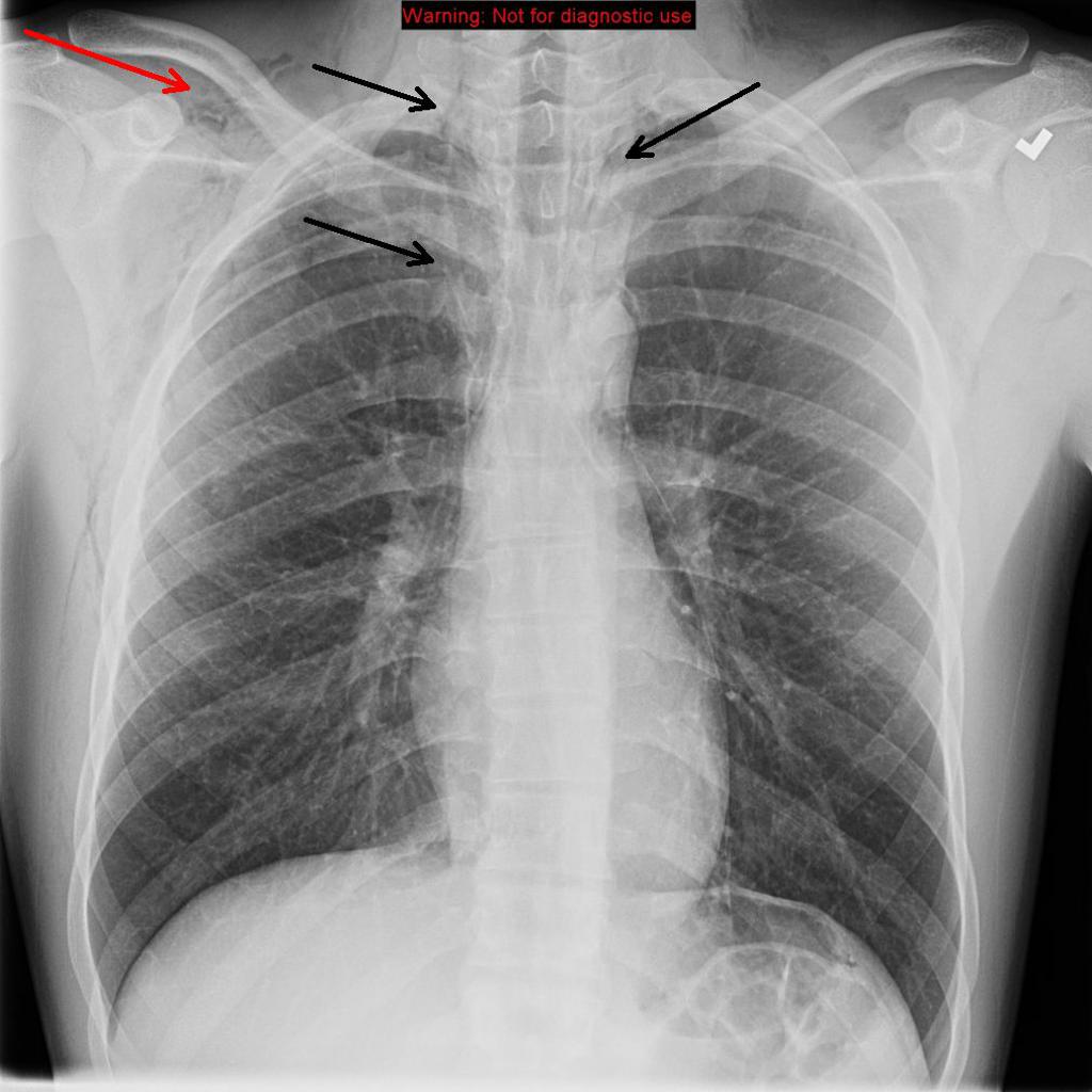 Chest X-ray: Frontal view reveals pneumomediastinum (black arrows). Subcutaneous emphysema (red arrow) along the chest wall, more prominent along the right than left; Source- Radiopaedia
 -
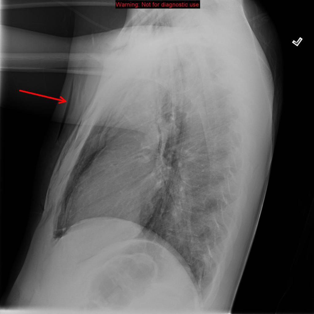 Chest X-ray: Boerhaave syndrome- Lateral radiographs subcutaneous emphysema (red arrow) along the chest wall; Source- Radiopaedia


References
References
- ↑ 1.0 1.1 Maurya VK, Sharma P, Ravikumar R, Bhatia M (2016). “Boerhaave’s syndrome”. Med J Armed Forces India. 72 (Suppl 1): S105–S107. doi:10.1016/j.mjafi.2015.12.004. PMC 5192176. PMID 28050085.
- ↑ 2.0 2.1 Tonolini M, Bianco R (2013). “Spontaneous esophageal perforation (Boerhaave syndrome): Diagnosis with CT-esophagography”. J Emerg Trauma Shock. 6 (1): 58–60. doi:10.4103/0974-2700.106329. PMC 3589863. PMID 23493470.
- ↑ Pate JW, Walker WA, Cole FH, Owen EW, Johnson WH (1989). “Spontaneous rupture of the esophagus: a 30-year experience”. Ann. Thorac. Surg. 47 (5): 689–92. PMID 2730190.
Editor-In-Chief: C. Michael Gibson, M.S., M.D. [1] Associate Editor(s)-in-Chief: Mohamed Diab, MD [2], Shaghayegh Habibi, M.D.[3], Ajay Gade MD[4]], Feham Tariq, MD [5]