Reliable, easy-to-read patient information on common medical conditions — written in plain language, free of jargon, and grounded in current evidence. Browse the most frequent diagnoses, understand what they mean for everyday life, and learn which steps and questions help you have a focused conversation with your doctor.
Synonyms and keywords: Wyburn mason’s syndrome; Retinoencephalofacial angiomatosis
Overview
Overview
Bonnet-Dechaume-Blanc syndrome or Wyburn mason’s syndrome or Retinoencephalofacial angiomatosis is a rare arteriovenous malformation (AVMs) condition. The special name for wyburn mason’s syndrome is called congenital retinocephalofacial vascular malformation syndrome” (“CRC syndrome”). In The United States of America in order to categorise a condition as a rare disease it should affect fewer than 200,000 people. Rare diseases also called as orphan diseases. Orphan Drug Act was passed on 1983 by congress for the rare diseases. Today an average of 25-30 million americans have been reported with rare diseases. The number of people with individual rare disease may be less but overall the number of people with rare diseases are large in number.
Historical Perspective
Historical Perspective
Bonnet-Dechaume-Blanc syndrome was first discovered by Magnus, in 1874.[1]
In 1932, Yates and Payne was the first to discover retinal and cerebral arteriovenous malformation (AVMs) in patients with Bonnet-Dechaume-Blanc syndrome.[2]
The exact pathogenesis of Bonnet-Dechaume-Blanc syndrome is not completely understood.
It is understood that Bonnet-Dechaume-Blanc syndrome may be caused by genetic factors but which are involved are difficult to say.
It is understood that Bonnet-Dechaume-Blanc syndrome is also may be caused by anomaly in organogenesis.
Origin of cells of the vessel walls in the cephalic region in the brain, and their migration may explain the connection between lesions looks alike but at different locations in the body.
Vision loss in patients with Bonnet-Dechaume-Blanc syndrome can be due to retinalarteriovenous malformations(AVM) which are tend to be large and lead to obscuration of the visual centers in the eye.
The cause of Bonnet-Dechaume-Blanc syndrome has not been identified yet.
Genetic Causes
Bonnet-Dechaume-Blanc syndrome may be is caused by a mutation in the genes.
Risk Factors
Risk Factors
There are no established risk factors for Bonnet-Dechaume-Blanc syndrome.
Natural History, Complications, and Prognosis
Natural History, Complications, and Prognosis
Natural History
The symptoms of Bonnet-Dechaume-Blanc syndrome usually develop in the first decade of life, and start with symptoms such as visual-field abnormalities.
Fundus picture of left eye with extended AVMs of the whole retina. The small arrows mean a slight increase of vascular loops. Case courtesy D Schmidt Et Al [18]Macular ischemia and extensive arteriovenous communications and dilated intertwined vessels. Case courtesy by Dimitrios A Karagiannis[19]
Incidence
Fewer than 100 cases of Bonnet-Dechaume-Blanc syndrome have been reported worldwide.[20]
The incidence of Bonnet-Dechaume-Blanc syndrome is unknown yet.
Prevalence
The prevalence of Bonnet-Dechaume-Blanc syndrome is unknown yet.
Digital subtraction angiography (DSA) of the left internal carotid artery: arterial phase (a, b) demonstrating the nidus of the arteriovenous malformation located around the chiasm (arrows) and behind the globe (arrowhead). In c venous drainage of the arteriovenous malformation and chorioretinal blush (open arrow). Case courtesy by D Schmidt Et Al[49]
Fundus photographs,fluorescein angiographic(FA)image, and enhanced depth imaging optical coherence tomographic(EDI-OCT) images. A:Markedly dilated tortuous vascular loops. The optic disc is obscured by very large vascular loops. Numerous anastomosing vessels make it difficult to separate the arterial and venous components. B: Fluorescein angiography in early phase shows fluorescein throughout the vascular loops without an intervening capillary bed and leakage from the loops, indicating a direct arteriovenous communication. Red arrow indicates the fovea (center of the foveal avascular zone), and green arrows indicate the direction of the OCT scans in ‘E’ and ‘F’. C:The vascular loops in some areas are less dilated and tortuous than in the left eye (see ‘A’). D–F: EDI-OCT images in the healthy right eye (D) and the affected left eye (E, F). Choroidal thickness of the left eye is thicker than that of the fellow eye. ‘E’ indicates a horizontal scan, and ‘D’ and ‘F’ indicate vertical scans through the fovea.(E, F) demonstrate retinal edema with cystic changes and oval-shaped lesions corresponding to cross sections of abnormal retinal vessels. White arrow indicates cystoid macular edema (F). Case courtesy by Akiko Iwata et Al.[50]
Head CT scan may be helpful in the diagnosis of Bonnet-Dechaume-Blanc syndrome. Findings on CT scan suggestive of Bonnet-Dechaume-Blanc syndrome include:
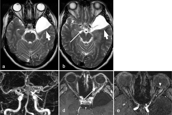
MRI showing a temporal arachnoidal cysttemporal arachnoidal cyst (short arrow in a, b), the enlarged retrobulbar vessels (long arrow in d), and retinal AVM (arrow head in e). MR-angiography in coronal view showing the chiasmal and hypothalamic network of the arterio-venous nidus (long arrow in c). Case courtesy by D Schmidt Et Al [57]Head MRI scan may be helpful in the diagnosis of Bonnet-Dechaume-Blanc syndrome. Findings on MRI scan suggestive of Bonnet-Dechaume-Blanc syndrome include:[58][59][60]
The majority of patients with Bonnet-Dechaume-Blanc syndrome shows no symptoms with unruptured arteriovenous malformations and these patients are under observation.[62]
↑Magnus, Hugo (1874). “Aneurysma arterioso-venosum retinale”. Archiv für Pathologische Anatomie und Physiologie und für Klinische Medicin. 60 (1): 38–45. doi:10.1007/BF01938766. ISSN0945-6317.
↑Yates, A. Gurney; Paine, C. G. (1930). “A CASE OF ARTERIOVENOUS ANEURYSM WITHIN THE BRAIN”. Brain. 53 (1): 38–46. doi:10.1093/brain/53.1.38. ISSN0006-8950.
↑Bhattacharya, J.J.; Luo, C.B.; Suh, D.C.; Alvarez, H.; Rodesch, G.; Lasjaunias, P. (2016). “Wyburn-Mason or Bonnet-Dechaume-Blanc as Cerebrofacial Arteriovenous Metameric Syndromes (CAMS)”. Interventional Neuroradiology. 7 (1): 5–17. doi:10.1177/159101990100700101. ISSN1591-0199.
↑Wyburn-Mason, R. (1943). “ARTERIOVENOUS ANEURYSM OF MID-BRAIN AND RETINA, FACIAL NÆVI AND MENTAL CHANGES”. Brain. 66 (3): 163–203. doi:10.1093/brain/66.3.163. ISSN0006-8950.
↑Dens, Helena; Casteels, Ingele (2018). “Exudative Type 3 Retinal Arteriovenous Malformation in a Pediatric Patient”. Case Reports in Ophthalmology. 9 (3): 504–509. doi:10.1159/000495656. ISSN1663-2699.
↑Callahan AB, Skondra D, Krzystolik M, Yonekawa Y, Eliott D (2015). “Wyburn-Mason Syndrome Associated With Cutaneous Reactive Angiomatosis and Central Retinal Vein Occlusion”. Ophthalmic Surg Lasers Imaging Retina. 46 (7): 760–2. doi:10.3928/23258160-20150730-12. PMID26247458.
↑Onder HI, Alisan S, Tunc M (March 2015). “Serous retinal detachment and cystoid macular edema in a patient with Wyburn-Mason syndrome”. Semin Ophthalmol. 30 (2): 154–6. doi:10.3109/08820538.2013.835832. PMID24171831.
↑Hon, Jennifer M.L.; Bhattacharya, Jo J.; Counsell, Carl E.; Papanastassiou, Vakis; Ritchie, Vaughn; Roberts, Richard C.; Sellar, Robin J.; Warlow, Charles P.; Al-Shahi Salman, Rustam (2009). “The Presentation and Clinical Course of Intracranial Developmental Venous Anomalies in Adults”. Stroke. 40 (6): 1980–1985. doi:10.1161/STROKEAHA.108.533034. ISSN0039-2499.
↑Hon, Jennifer M.L.; Bhattacharya, Jo J.; Counsell, Carl E.; Papanastassiou, Vakis; Ritchie, Vaughn; Roberts, Richard C.; Sellar, Robin J.; Warlow, Charles P.; Al-Shahi Salman, Rustam (2009). “The Presentation and Clinical Course of Intracranial Developmental Venous Anomalies in Adults”. Stroke. 40 (6): 1980–1985. doi:10.1161/STROKEAHA.108.533034. ISSN0039-2499.
↑Bloom, P. A; Laidlaw, A.; Easty, D. L (1993). “Spontaneous development of retinal ischaemia and rubeosis in eyes with retinal racemose angioma”. British Journal of Ophthalmology. 77 (2): 124–125. doi:10.1136/bjo.77.2.124. ISSN0007-1161.
↑Medina FM, Maia OO, Takahashi WY (2010). “Rhegmatogenous retinal detachment in Wyburn-Mason syndrome: case report”. Arq Bras Oftalmol. 73 (1): 88–91. PMID20464122.
↑Schmidt D, Pache M, Schumacher M (2008). “The congenital unilateral retinocephalic vascular malformation syndrome (bonnet-dechaume-blanc syndrome or wyburn-mason syndrome): review of the literature”. Surv Ophthalmol. 53 (3): 227–49. doi:10.1016/j.survophthal.2007.10.001. PMID18501269.
↑Meyer, Carsten H.; Rodrigues, Eduardo B.; Mennel, Stefan; Klingmüller, Volker; Kroll, Peter (2007). “Functional and anatomical investigations in racemose haemangioma”. Acta Ophthalmologica Scandinavica. 85 (7): 764–771. doi:10.1111/j.1600-0420.2007.00911.x. ISSN1395-3907.
↑Augsburger JJ, Goldberg RE, Shields JA, Mulberger RD, Magargal LE (1980). “Changing appearance of retinal arteriovenous malformation”. Albrecht Von Graefes Arch Klin Exp Ophthalmol. 215 (1): 65–70.
↑Zeng Y, Fan YC, Liu Y, Wan L (April 2016). “Cerebral Arteriovenous Malformation in Wyburn-Mason Syndrome”. J Pediatr Ophthalmol Strabismus. 53 Online: e15–7. doi:10.3928/01913913-20160406-01. PMID27112169.
↑Karagiannis, Dimitrios; Panagiotidis, Dimitrios; Tsoubris, Ioannis; Theodossiadis, Panagiotis (2011). “Spontaneous development of macular ischemia in a case of racemose hemangioma”. Clinical Ophthalmology: 931. doi:10.2147/OPTH.S21925. ISSN1177-5483.
↑Fujita H, Nakano K, Kumon Y, Inoue H, Sakaki S (August 1989). “[A case of Wyburn-Mason syndrome]”. Rinsho Shinkeigaku (in Japanese). 29 (8): 1039–44. PMID2598528.CS1 maint: Unrecognized language (link)
↑Zeng Y, Fan YC, Liu Y, Wan L (April 2016). “Cerebral Arteriovenous Malformation in Wyburn-Mason Syndrome”. J Pediatr Ophthalmol Strabismus. 53 Online: e15–7. doi:10.3928/01913913-20160406-01. PMID27112169.
↑Archer, Desmond B.; Deutman, August; Ernest, J. Terry; Krill, Alex E. (1973). “Arteriovenous Communications of the Retina”. American Journal of Ophthalmology. 75 (2): 224–241. doi:10.1016/0002-9394(73)91018-0. ISSN0002-9394.
↑Onder HI, Alisan S, Tunc M (March 2015). “Serous retinal detachment and cystoid macular edema in a patient with Wyburn-Mason syndrome”. Semin Ophthalmol. 30 (2): 154–6. doi:10.3109/08820538.2013.835832. PMID24171831.
↑Iwata, Akiko; Mitamura, Yoshinori; Niki, Masanori; Semba, Kentaro; Egawa, Mariko; Katome, Takashi; Sonoda, Shozo; Sakamoto, Taiji (2015). “Binarization of enhanced depth imaging optical coherence tomographic images of an eye with Wyburn-Mason syndrome: a case report”. BMC Ophthalmology. 15 (1). doi:10.1186/s12886-015-0014-2. ISSN1471-2415.
↑Kittelberger, Reinhold; Davis, Paul F.; Stehbens, William E. (1989). “An improved immunofluorescence techniquefor the histological examination of blood vessel tissue”. Acta Histochemica. 86 (2): 137–IN2. doi:10.1016/S0065-1281(89)80082-0. ISSN0065-1281.
↑Schmidt, D; Agostini, H; Schumacher, M (2010). “Twenty-seven years follow-up of a patient with congenital retinocephalofacial vascular malformation syndrome and additional congenital malformations (Bonnet-dechaume-blanc syndrome or wyburn-mason syndrome)”. European Journal of Medical Research. 15 (2): 88. doi:10.1186/2047-783X-15-2-88. ISSN2047-783X.
↑Schmidt, D; Agostini, H; Schumacher, M (2010). “Twenty-seven years follow-up of a patient with congenital retinocephalofacial vascular malformation syndrome and additional congenital malformations (Bonnet-dechaume-blanc syndrome or wyburn-mason syndrome)”. European Journal of Medical Research. 15 (2): 88. doi:10.1186/2047-783X-15-2-88. ISSN2047-783X.
↑Numaguchi Y, Kitamura K, Fukui M, Ikeda J, Hasuo K, Kishikawa T; et al. (1982). “Intracranial venous angiomas”. Surg Neurol. 18 (3): 193–202. PMID7179074.CS1 maint: Explicit use of et al. (link) CS1 maint: Multiple names: authors list (link)
↑Dayani PN, Sadun AA (May 2007). “A case report of Wyburn-Mason syndrome and review of the literature”. Neuroradiology. 49 (5): 445–56. doi:10.1007/s00234-006-0205-x. PMID17235577.
↑Slim E, Antoun J, Kourie HR, Schakkal A, Cherfan G (October 2014). “Intravitreal bevacizumab for retinal capillary hemangioblastoma: A case series and literature review”. Can. J. Ophthalmol. 49 (5): 450–7. doi:10.1016/j.jcjo.2014.07.007. PMID25284102.
↑Chatziralli, Irini; Maniatea, Aggeliki; Koubouni, Kyriakoula; Parikakis, Efstratios; Mitropoulos, Panagiotis (2016). “Intravitreal Ranibizumab for Retinal Arterial Macroaneurysm: Long-Term Results of a Prospective Study”. European Journal of Ophthalmology. 27 (2): 215–219. doi:10.5301/ejo.5000863. ISSN1120-6721.
↑Bormann, Caroline; Heichel, Jens; Hammer, Ute; Habermann, Anke; Hammer, Thomas (2017). “Intravitreal Anti-Vascular Endothelial Growth Factor for Macular Edema due to Complex Retinal Arterial Macroaneurysms”. Case Reports in Ophthalmology. 8 (1): 141–147. doi:10.1159/000458517. ISSN1663-2699.
↑Lavin, M J; Marsh, R J; Peart, S; Rehman, A (1987). “Retinal arterial macroaneurysms: a retrospective study of 40 patients”. British Journal of Ophthalmology. 71 (11): 817–825. doi:10.1136/bjo.71.11.817. ISSN0007-1161.
↑Abdel-Khalek, M. N.; Richardson, J. (1986). “Retinal macroaneurysm: natural history and guidelines for treatment”. British Journal of Ophthalmology. 70 (1): 2–11. doi:10.1136/bjo.70.1.2. ISSN0007-1161.