
Bronchopulmonary segment
Editor-In-Chief: C. Michael Gibson, M.S., M.D. [1]
Each of the tertiary bronchi serves a specific bronchopulmonary segment. These segments each have their own artery. Thus, each bronchopulmonary segment is supplied by a bronchus, and an artery.
There are 10 bronchopulmonary segments per lung (3 in right superior lobe, 2 in middle right lobe, 5 in both right and left inferior lobe, and 5 in left superior lobe, in which 2 of the five reside in the lingula), each of which is separated from the others by a layer of connective tissue.
This means that each bronchopulmonary segment is a discrete anatomical and functional unit, and this separation mean that a bronchopulmonary segment can be surgically removed without affecting the function of the other segments.
Bronchopulmonary segments of right lung
Bronchopulmonary segments of right lung
A PALM Seed Makes Another Little Palm
- superior lobe
- apical
- posterior
- anterior
- middle lobe
- lateral (superior lingular)
- medial (inferior lingular)
- inferior lobe
- superior
- medial-basal
- anterior-basal
- lateral-basal
- posterior-basal
Editor-In-Chief: C. Michael Gibson, M.S., M.D. [1]
Overview
The Right lung is divided into three lobes, superior, middle, and inferior, by two interlobular fissures:
Fissures
- One of these, the oblique fissure, separates the inferior from the middle and superior lobes, and corresponds closely with the fissure in the left lung. Its direction is, however, more vertical, and it cuts the lower border about 7.5 cm. behind its anterior extremity.
- The other fissure, the horizontal fissure, separates the superior from the middle lobe. It begins in the previous fissure near the posterior border of the lung, and, running horizontally forward, cuts the anterior border on a level with the sternal end of the fourth costal cartilage; on the mediastinal surface it may be traced backward to the hilus.
Lobes
The middle lobe, the smallest lobe of the right lung, is wedge-shaped, and includes the lower part of the anterior border and the anterior part of the base of the lung. (There is no middle lobe on the left lung, though there is a lingula.)
The superior and inferior lobes are similar to their counterparts on the left lung.
Difference in size
The right lung, although shorter by 2.5 cm. than the left, in consequence of the diaphragm rising higher on the right side to accommodate the liver, is broader, owing to the inclination of the heart to the left side; its total capacity is greater and it weighs more than the left lung.
Impressions
On the mediastinal surface, immediately above the hilus, is an arched furrow which accommodates the azygos vein; while running upward, and then arching lateralward some little distance below the apex, is a wide groove for the superior vena cava and right innominate vein; behind this, and nearer the apex, is a furrow for the innominate artery.
Behind the hilus and the attachment of the pulmonary ligament is a vertical groove for the esophagus; this groove becomes less distinct below, owing to the inclination of the lower part of the esophagus to the left of the middle line.
In front and to the right of the lower part of the esophageal groove is a deep concavity for the extrapericardiac portion of the thoracic part of the inferior vena cava.
Additional images
-
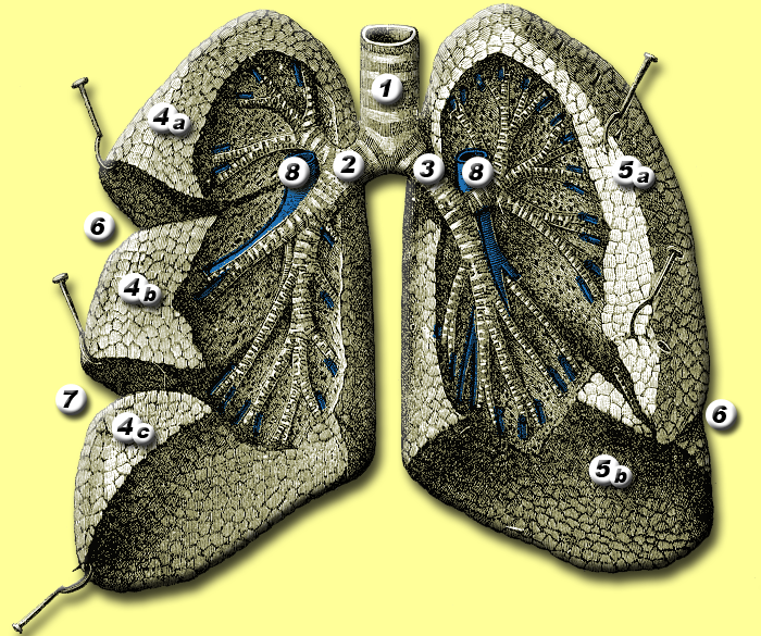 Anatomy of lungs.
 -
 Front view of heart and lungs.
 -
 Transverse section of thorax, showing relations of pulmonary artery.
 -
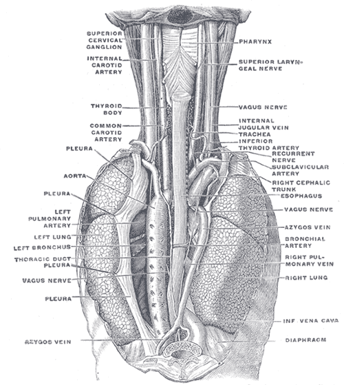 The position and relation of the esophagus in the cervical region and in the posterior mediastinum. Seen from behind.
 -
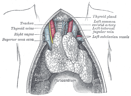 The thymus of a full-time fetus, exposed in situ.


See also
External links
- Lung Lobes
- Template:Chorus
- Template:SUNYAnatomyFigs – “Mediastinal surface of the right lung.”
- Diagram and quiz at cancer.gov
- Template:RocheLexicon
- Cross section at univie.ac.at
Bronchopulmonary segments of left lung
Bronchopulmonary segments of left lung
ASIA ALPS
Apoptotic Antlions Stop In, Suddenly Amalgamating Laboratory Posts
AP And Supine alignment Increases Limited Studies And Makes Baseline Pulmonary Bases Look Bad (a radiology mnemonic)
- superior lobe
- apico-posterior (merger of “apical” and “posterior”)
- anterior
- lingula of superior lobe
- inferior lingular
- superior lingular
- inferior lobe
- superior
- antero-medial basal (merger of “anterior basal” and “medial basal”)
- lateral basal
- posterior basal
External links
External links
- Template:EMedicineDictionary
- Template:GPnotebook
- Photo at ccccd.edu
- Template:SUNYAnatomyLabs – “Pleural Cavities and Lungs: Bronchopulmonary segments”
- Illustration at uams.edu
- Sealy W, Connally S, Dalton M (1993). “Naming the bronchopulmonary segments and the development of pulmonary surgery”. Ann Thorac Surg. 55 (1): 184–8. PMID 8417676.
- Template:MedicalMnemonics
Looking for the patient version?
© 2026 MyEClinic – IFTM Institut für Telematik in der Medizin GmbH