
Bulbus cordis
Editor-In-Chief: C. Michael Gibson, M.S., M.D. [1]
Overview
Overview
When the developing heart assumes its S-shaped form, the bulbus cordis lies ventral to the primitive ventricle. Together, the bulbus cordis and the primitive ventricle give rise to the ventricle of the formed heart.
The adjacent walls of the bulbus cordis and ventricle approximate, fuse, and finally disappear, and the bulbus cordis now communicates freely with the right ventricle, while the junction of the bulbus with the truncus arteriosus is brought directly ventral to and applied to the atrial canal.
By the upgrowth of the ventricular septum the bulbus cordis is in great measure separated from the left ventricle, but remains an integral part of the right ventricle, of which it forms the infundibulum.
Additional images
Additional images
-
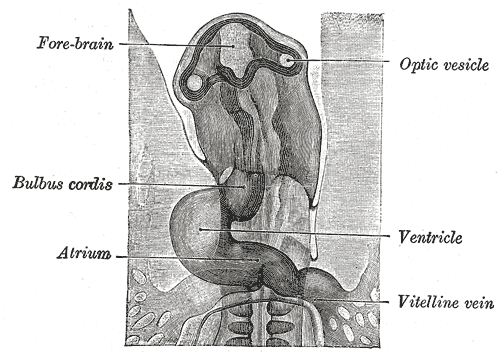 Head of chick embryo of about thirty-eight hours’ incubation, viewed from the ventral surface. X 26
 -
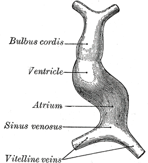 Diagram to illustrate the simple tubular condition of the heart.
 -
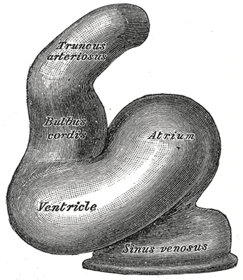 Heart of human embryo of about fourteen days.
 -
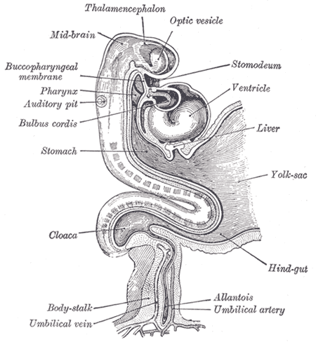 Human embryo about fifteen days old. Brain and heart represented from right side. Digestive tube and yolk sac in median section.


Looking for the patient version?
© 2026 MyEClinic – IFTM Institut für Telematik in der Medizin GmbH