

Health Dictionary
Reliable, easy-to-read patient information on common medical conditions — written in plain language, free of jargon, and grounded in current evidence. Browse the most frequent diagnoses, understand what they mean for everyday life, and learn which steps and questions help you have a focused conversation with your doctor.
Burkitt's lymphoma other imaging studies
Introduction
Other Imaging Studies
Other Imaging Studies
PET and bone scan may be helpful in the diagnosis of Burkitt’s lymphoma.[1]
PET scan
The following finding on PET scan may be suggestive of Burkitt’s lymphoma:
- [18F]-2-deoxy-D-glucose uptake in affected nodes or tissue[2]
Ultrasound
The following finding on ultrasound may be suggestive of Burkitt’s lymphoma:
- Thickening of organs
- Free fluid [3]
- Intussusception[4]

Bone scan
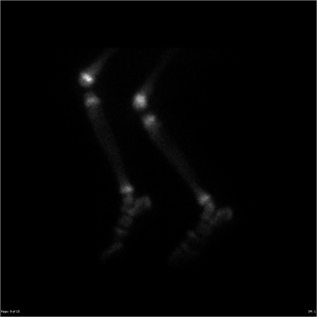
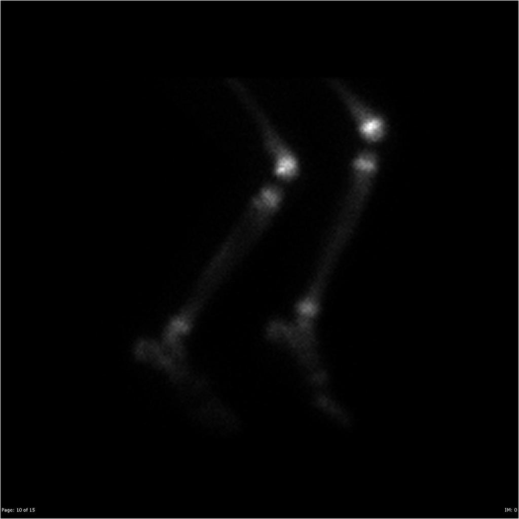
The following finding on bone scan may be suggestive of Burkitt’s lymphoma:
-
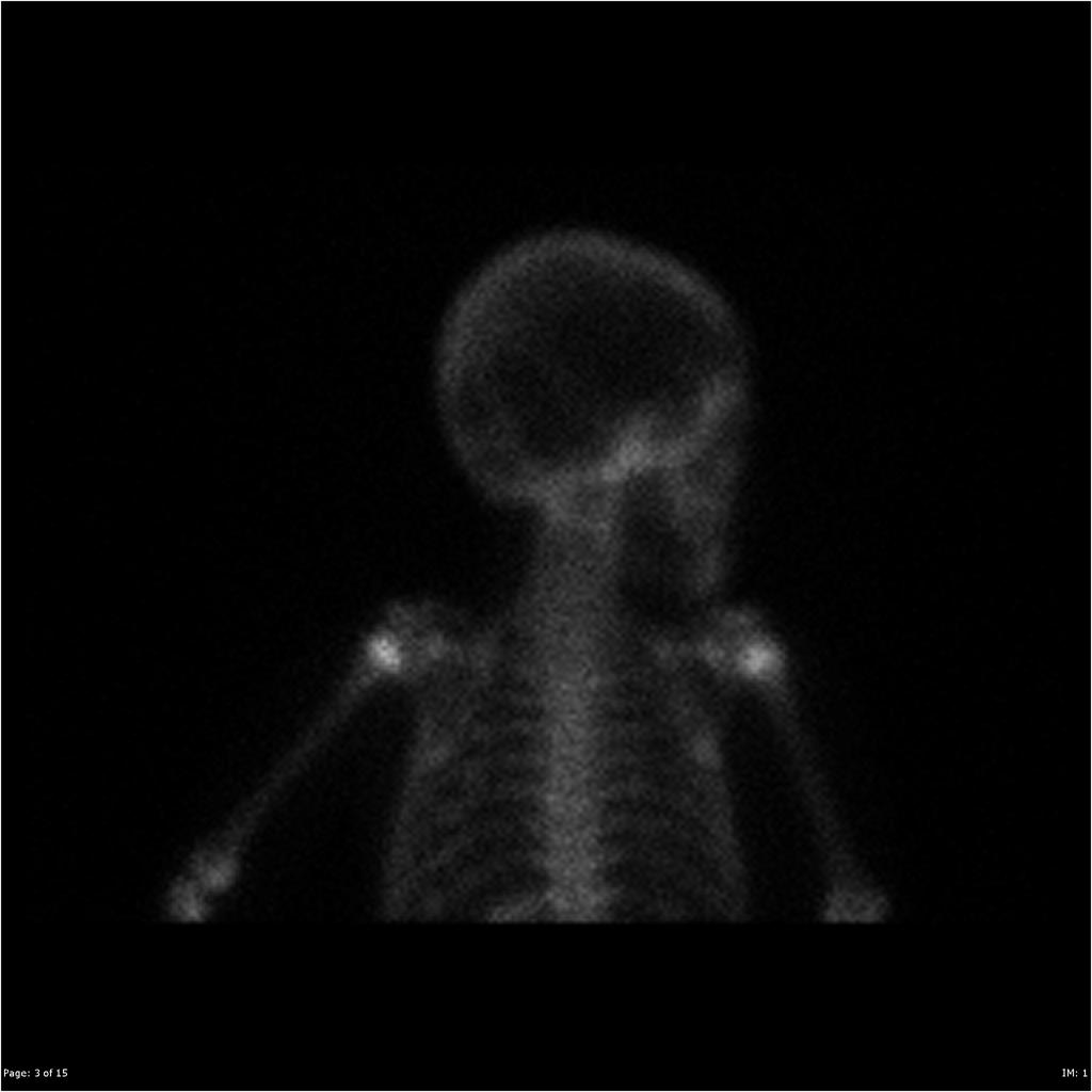 A
A -
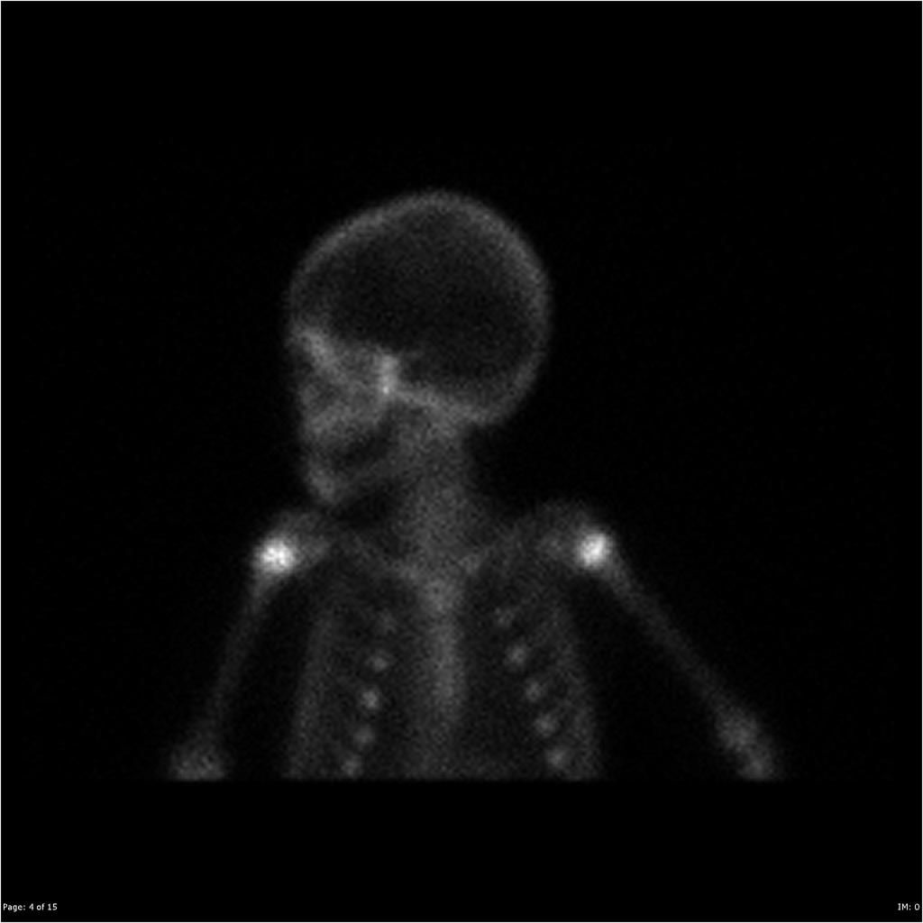 B
B -
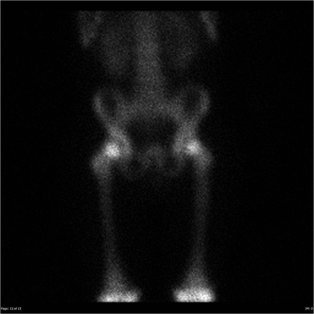 C
C -
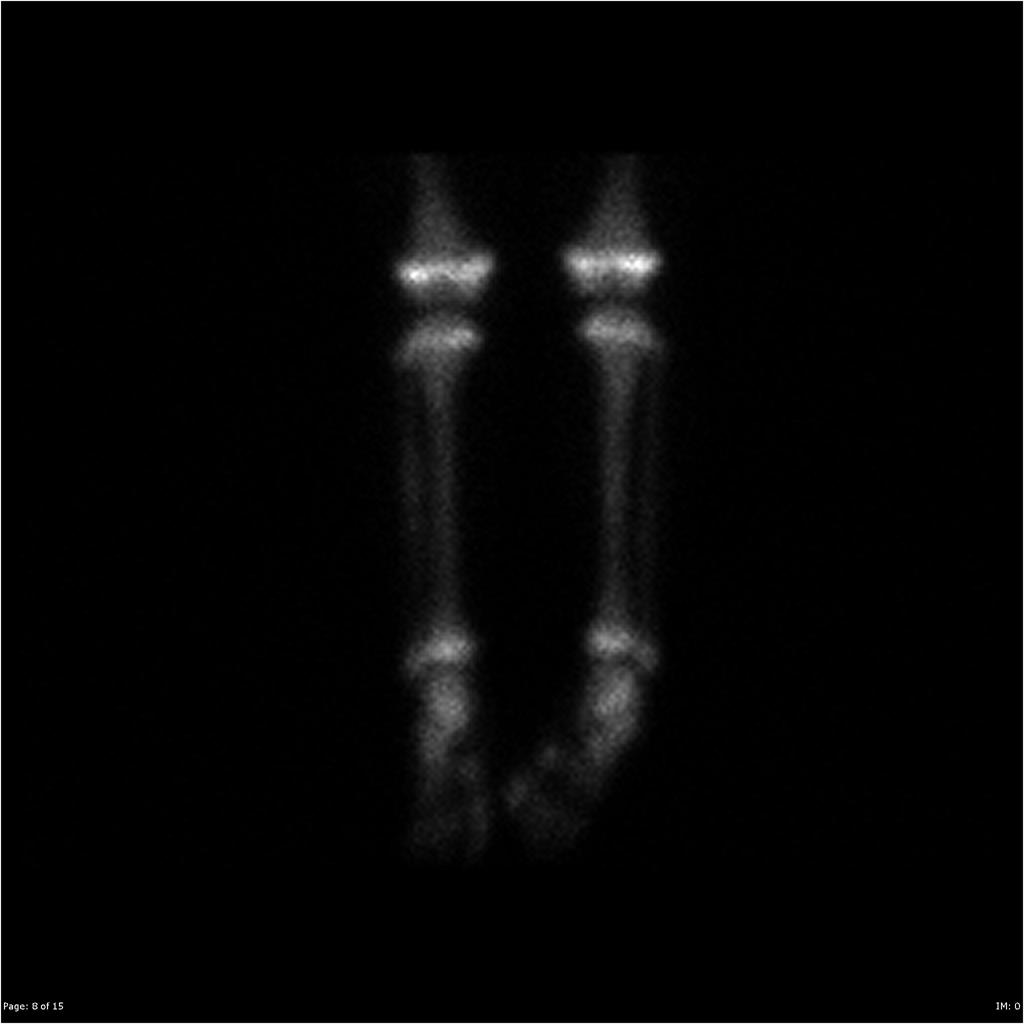 D
D -
 E
E -
 F
F


- Figures A-F: demonstrate a global increase in contrast metaphyseal uptake[5]
References
References
- ↑ Burkitt lymphoma. MedlinePlus. https://www.nlm.nih.gov/medlineplus/ency/article/001308.htm Accessed on September 30, 2015
- ↑ Carrillo-Cruz E, Marín-Oyaga VA, Solé Rodríguez M, Borrego-Dorado I, de la Cruz Vicente F, Quiroga Cantero E; et al. (2015). “Role of 18F-FDG-PET/CT in the management of Burkitt lymphoma”. Eur J Haematol. 94 (1): 23–30. doi:10.1111/ejh.12284. PMID 24520874.
- ↑ Bernardi D, Asti E, Bonavina L (2016). “Adult ileocolic intussusception caused by Burkitt lymphoma”. BMJ Case Rep. 2016. doi:10.1136/bcr-2016-218334. PMC 5174892. PMID 27920022.
- ↑ Gale HI, Gee MS, Westra SJ, Nimkin K (2016). “Abdominal ultrasonography of the pediatric gastrointestinal tract”. World J Radiol. 8 (7): 656–67. doi:10.4329/wjr.v8.i7.656. PMC 4965350. PMID 27551336.
- ↑ Image courtesy of Dr Jeremy Jones. Radiopaedia(original file ‘’here’’).Creative Commons BY-SA-NC
Editor-In-Chief: C. Michael Gibson, M.S., M.D. [1]; Associate Editor(s)-in-Chief: Sowminya Arikapudi, M.B,B.S. [2], Kamal Akbar, M.D.[3]