

Health Dictionary
Reliable, easy-to-read patient information on common medical conditions — written in plain language, free of jargon, and grounded in current evidence. Browse the most frequent diagnoses, understand what they mean for everyday life, and learn which steps and questions help you have a focused conversation with your doctor.
Calyceal diverticulum
Introduction
Synonyms and Related Keywords: Pyelocalyceal diverticulum
Overview
Overview
Calyceal diverticulum is a relatively uncommon cyst-like lesion, often discovered incidentally during ultrasound examination of the kidneys. Calyceal diverticulum is a urine-containing cavity within the renal parenchyma, communicating with the collecting system through a narrow channel. [1]
Classification
Classification
- Two types are identified:
- One is related to a minor calyx and usually located in the upper pole
- Other is connected with the pelvis or a major calyx in the central portion of the kidney.
- The majority of calyceal diverticulum are small and asymptomatic, and do not require any treatment.
- Calyceal diverticulua may rarely cause urinary tract infection, renal colic, pyuria, hematuria or hypertension.
- Mobile calculi and milk of calcium are characteristic findings in calyceal diverticulum.
Diagnosis
Diagnosis
Ultrasound
- Ultrasound examination suggests the diagnosis of calyceal diverticulum and it has a variable appearance.
- The diverticulum may appear as a cyst-like lesion, indistinguishable from a simple cyst.
- Recognition of mobile echogenic material within such a cyst-like structure is considered characteristic of milk of calcium. A cyst-like mass with milk of calcium is presumed to be a calyceal diverticulum.
CT
- CT has an important role in the presence of a complicated cystic mass diagnosed by ultrasound without the characteristic features of calyceal diverticulum.
- The demonstration of the layering of contrast medium on delayed images is pathognomonic of these lesions.
- The patency of the diverticulum is demonstrated by the gradual opacification on delayed images.
-
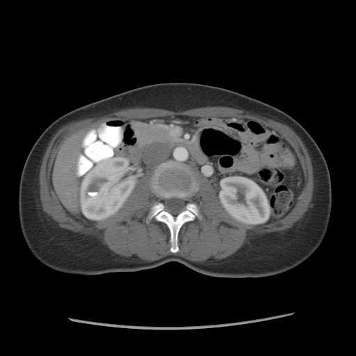 CT: Calyceal diverticulum
 -
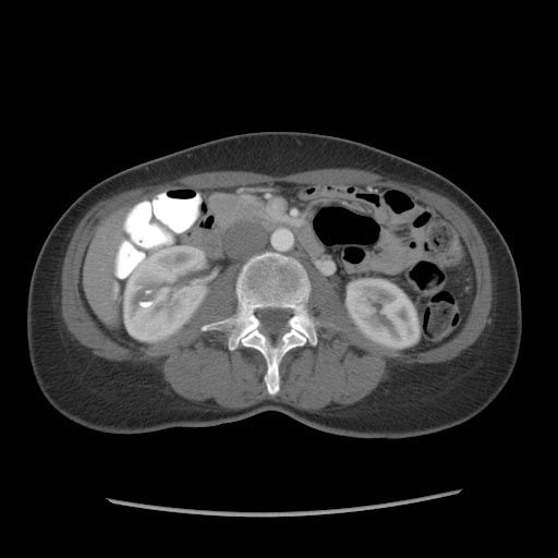 CT: Calyceal diverticulum


References
References
- ↑ Rathaus, V, Konen, O, Werner, M, Shapiro Feinberg, M, Grunebaum, M, Zissin, R. Pyelocalyceal diverticulum: the imaging spectrum with emphasis on the ultrasound features. Br J Radiol 2001 74: 595-601.
Editor-In-Chief: C. Michael Gibson, M.S., M.D. [1] Associate Editor(s)-in-Chief: Cafer Zorkun M.D., PhD.