

Health Dictionary
Reliable, easy-to-read patient information on common medical conditions — written in plain language, free of jargon, and grounded in current evidence. Browse the most frequent diagnoses, understand what they mean for everyday life, and learn which steps and questions help you have a focused conversation with your doctor.
Campylobacteriosis causes
Introduction
Overview
Overview
Campylobacter jejuni is a species of curved,long rod-shaped, Gram-negative microaerophilic, bacteria commonly found in animal feces.[1] It is one of the most common causes of human gastroenteritis in the world. Food poisoning caused by Campylobacter species can be severely debilitating but is rarely life-threatening. It has been linked with subsequent development of Guillain-Barré syndrome (GBS), which usually develops two to three weeks after the initial illness.
Organism
Organism
C. jejuni is commonly associated with poultry and naturally colonises the GI tract of many bird species. It has also been isolated from wombat and kangaroo feces, being a cause of bushwalkers’ diarrhea. Contaminated drinking water and unpasteurized milk provide an efficient means for distribution. Contaminated food is a major source of isolated infections, with incorrectly prepared meat and raw poultry normally the source of the bacteria.
Infection with C. jejuni usually results in enteritis, which is characterised by abdominal pain, diarrhea, fever, and malaise. The symptoms usually persist for between 24 hours and a week, but may be longer. Diarrhea can vary in severity from loose stools to bloody stools. No antibiotics are usually given as the disease is self-limiting, however, severe or prolonged cases may require ciprofloxacin, erythromycin or norfloxacin. Fluid and electrolyte replacement may be required for serious cases.
Laboratory characteristics
Laboratory characteristics
| Characteristic | Result |
|---|---|
| Growth at 25 °C | – |
| Growth at 35-37 °C | – |
| Growth at 42 °C | + |
| Nitrate reduction | + |
| Catalase test | + |
| Oxidase test | + |
| Growth on MacConkey agar | + |
| Motility (wet mount) | + |
| Glucose utilization | – |
| Hippurate hydrolysis | + |
| Resistance to naladixic acid | – |
| Resistance to cephalothin | + |
Campylobacter is grown on specially selective “CAMP” agar plates at 42 °C, the normal avian body temperature, rather than at 37 °C, the temperature at which most other pathogenic bacteria are grown. Since the colonies are oxidase positive, they will usually only grow in scanty amounts on the plates. Microaerophilic conditions are required for luxurious growth. The selective medium known as Skirrow’s medium is used. Skirrow’s medium is blood agar infused with a cocktail of antibiotics: vancomycin, polymixin-B and trimethoprim under microaerophilic conditions at 42 degrees.
Gallery
Gallery
-
![Campylobacter fetus (C. fetus ss. jejuni) cultures were grown on Skirrow's and Butzler's medium. From Public Health Image Library (PHIL). [2]](https://www.wikidoc.org/images/1/19/Campylobacter12.jpeg) Campylobacter fetus (C. fetus ss. jejuni) cultures were grown on Skirrow’s and Butzler’s medium. From Public Health Image Library (PHIL). [2]
Campylobacter fetus (C. fetus ss. jejuni) cultures were grown on Skirrow’s and Butzler’s medium. From Public Health Image Library (PHIL). [2] -
![Campylobacter jejunii, formerly known as C. fetus subsp. jejuni, culture was grown on Skirrow's and Butzler's medium. From Public Health Image Library (PHIL). [2]](https://www.wikidoc.org/images/e/ee/Campylobacter11.jpeg) Campylobacter jejunii, formerly known as C. fetus subsp. jejuni, culture was grown on Skirrow’s and Butzler’s medium. From Public Health Image Library (PHIL). [2]
Campylobacter jejunii, formerly known as C. fetus subsp. jejuni, culture was grown on Skirrow’s and Butzler’s medium. From Public Health Image Library (PHIL). [2] -
![Scanning electron micrograph depicts a number of Gram-negative Campylobacter jejuni bacteria (11,734x mag). From Public Health Image Library (PHIL). [2]](https://www.wikidoc.org/images/f/f4/Campylobacter09.jpeg) Scanning electron micrograph depicts a number of Gram-negative Campylobacter jejuni bacteria (11,734x mag). From Public Health Image Library (PHIL). [2]
Scanning electron micrograph depicts a number of Gram-negative Campylobacter jejuni bacteria (11,734x mag). From Public Health Image Library (PHIL). [2] -
![Scanning electron micrograph depicts a number of Gram-negative Campylobacter jejuni bacteria (9,951x mag). From Public Health Image Library (PHIL). [2]](https://www.wikidoc.org/images/7/7f/Campylobacter07.jpeg) Scanning electron micrograph depicts a number of Gram-negative Campylobacter jejuni bacteria (9,951x mag). From Public Health Image Library (PHIL). [2]
Scanning electron micrograph depicts a number of Gram-negative Campylobacter jejuni bacteria (9,951x mag). From Public Health Image Library (PHIL). [2] -
![Micrograph depicts a 0.1µm polycarbonate membrane filter, which is coated with a C. jejuni bacterial film. From Public Health Image Library (PHIL). [2]](https://www.wikidoc.org/images/1/1c/Campylobacter06.jpeg) Micrograph depicts a 0.1µm polycarbonate membrane filter, which is coated with a C. jejuni bacterial film. From Public Health Image Library (PHIL). [2]
Micrograph depicts a 0.1µm polycarbonate membrane filter, which is coated with a C. jejuni bacterial film. From Public Health Image Library (PHIL). [2] -
![Campylobacter jejuni bacteria revealing characteristic “thin”, “comma-“, “S-“ or “gull winged-shaped” forms. From Public Health Image Library (PHIL). [2]](https://www.wikidoc.org/images/8/89/Campylobacter04.jpeg) Campylobacter jejuni bacteria revealing characteristic “thin”, “comma-“, “S-“ or “gull winged-shaped” forms. From Public Health Image Library (PHIL). [2]
Campylobacter jejuni bacteria revealing characteristic “thin”, “comma-“, “S-“ or “gull winged-shaped” forms. From Public Health Image Library (PHIL). [2] -
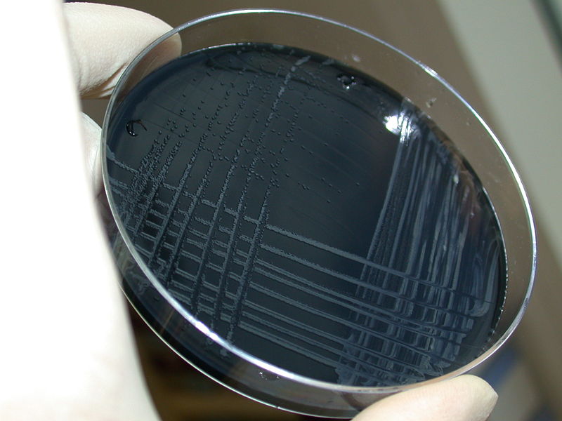 Blood-free, charcoal-based selective medium agar (CSM) for isolation of Campylobacter jejuni.
 -
Scanning electron micrograph depicting a number of Campylobacter jejuni bacteria.
![Campylobacter fetus (C. fetus ss. jejuni) cultures were grown on Skirrow's and Butzler's medium. From Public Health Image Library (PHIL). [2]](https://www.wikidoc.org/index.php/File%3ACampylobacter12.jpeg)
![Campylobacter jejunii, formerly known as C. fetus subsp. jejuni, culture was grown on Skirrow's and Butzler's medium. From Public Health Image Library (PHIL). [2]](https://www.wikidoc.org/index.php/File%3ACampylobacter11.jpeg)
![Scanning electron micrograph depicts a number of Gram-negative Campylobacter jejuni bacteria (11,734x mag). From Public Health Image Library (PHIL). [2]](https://www.wikidoc.org/index.php/File%3ACampylobacter09.jpeg)
![Scanning electron micrograph depicts a number of Gram-negative Campylobacter jejuni bacteria (9,951x mag). From Public Health Image Library (PHIL). [2]](https://www.wikidoc.org/index.php/File%3ACampylobacter07.jpeg)
![Micrograph depicts a 0.1µm polycarbonate membrane filter, which is coated with a C. jejuni bacterial film. From Public Health Image Library (PHIL). [2]](https://www.wikidoc.org/index.php/File%3ACampylobacter06.jpeg)
![Campylobacter jejuni bacteria revealing characteristic “thin”, “comma-“, “S-“ or “gull winged-shaped” forms. From Public Health Image Library (PHIL). [2]](https://www.wikidoc.org/index.php/File%3ACampylobacter04.jpeg)

References
References
- ↑ Ryan KJ, Ray CG (editors) (2004). Sherris Medical Microbiology (4th ed. ed.). McGraw Hill. ISBN 0-8385-8529-9.
- ↑ 2.0 2.1 2.2 2.3 2.4 2.5 “Public Health Image Library (PHIL)”.
- “Multiple Campylobacter Genomes Sequenced”. 2005-01-04. Retrieved 2007-07-27.
- Gorbach, Sherwood L., Falagas, Matthew (editors) (2001). The 5 minute infectious diseases consult (1st ed. ed.). Lippincott Williams & Wilkins. ISBN 0-683-30736-3.
- Online Bacteriological Analytical Manual, Chapter 7: Campylobacter – US Center for Food Safety & Applied Nutrition (accessed 13 October 2006)
Editor-In-Chief: C. Michael Gibson, M.S., M.D. [1]