
Cor triatriatum other imaging findings
Editor-In-Chief: C. Michael Gibson, M.S., M.D. [1]; Associate Editors-In-Chief: Priyamvada Singh, M.B.B.S. [2]; Cafer Zorkun, M.D., Ph.D. [3]; Keri Shafer, M.D. [4]; Assistant Editor(s)-In-Chief: Kristin Feeney, B.S. [5]
Overview
Overview
During diagnosis, additional methods of imaging may be used to better identify the nature of the cor triatriatum defect and its implication on cardiac blood flow. Below are imaging findings obtained through academic study of cor triatriatum.
Images
Images
Images shown below are courtesy of Professor Peter Anderson DVM PhD and Published with permission © PEIR, University of Alabama at Birmingham, Department of Pathology
-
 Cor Triatriatum: Gross left ventricular outflow tract appears normal
 -
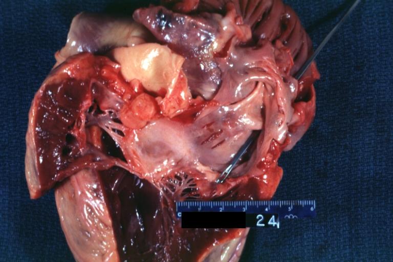
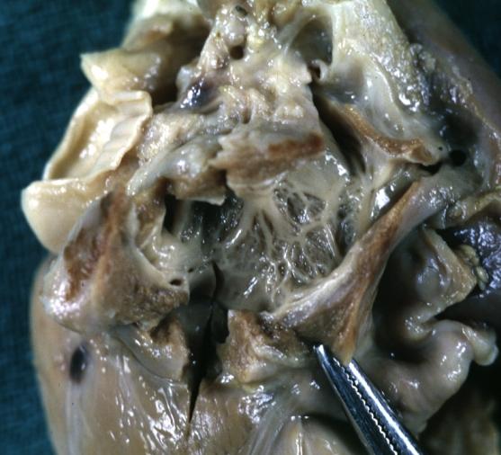
 Cor Triatriatum: Gross left atrial inlet chamber with probe extending through narrow connection to true left atrium below
 -
 Cor Triatriatum: Gross right atrium tricuspid valve and right ventricle note right ventricular hypertrophy
 -
 Cor Triatriatum: Gross
 -
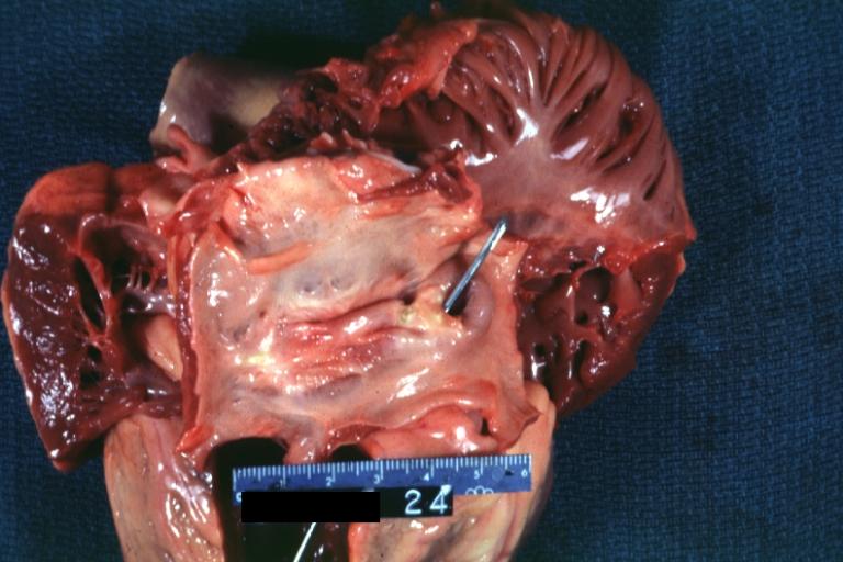
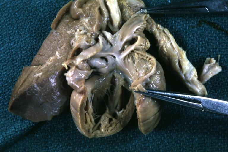
 Cor Triatriatum: Gross fixed tissue opened left atrium mitral valve and left ventricle
 -
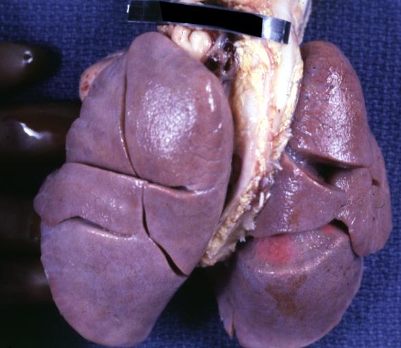 Lung: Abnormal Lobation: Gross fixed tissue posterior view both lungs with many lobes case of cor triatriatum
 -
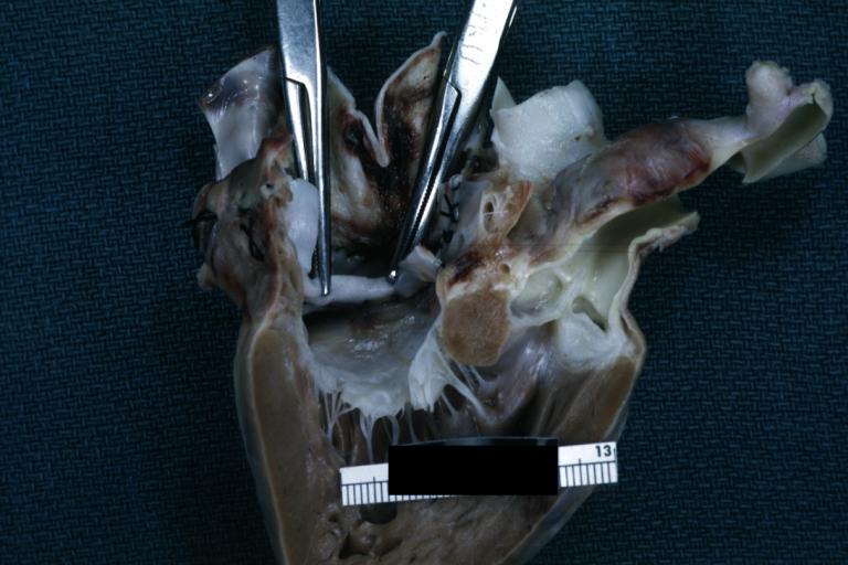
 Cor Triatriatum: Gross fixed tissue opened superior atrial chamber
 -
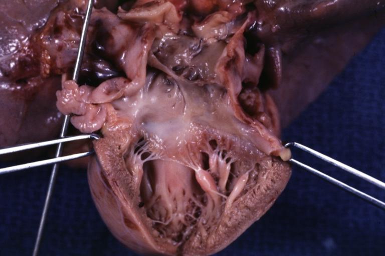
 Cor Triatriatum: Gross fixed tissue opened infant heart with the two chambered left atrium


Looking for the patient version?
© 2026 MyEClinic – IFTM Institut für Telematik in der Medizin GmbH