
Descending colon
Overview
Overview
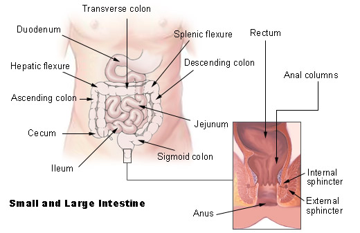
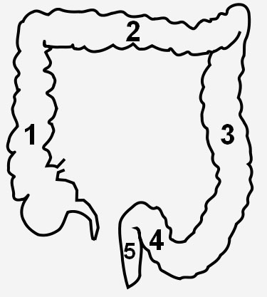
The descending colon of humans passes downward through the left hypochondrium and lumbar regions, along the lateral border of the left kidney.
At the lower end of the kidney it turns medialward toward the lateral border of the psoas muscle, and then descends, in the angle between psoas and quadratus lumborum, to the crest of the ilium, where it ends in the sigmoid colon.
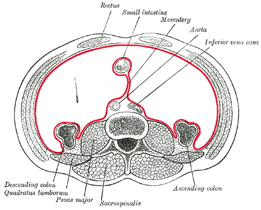
The peritoneum covers its anterior surface and sides, and therefore the descending colon is described as retroperitoneal. (The transverse colon and sigmoid colon, which are immediately proximal and distal, are intraperitoneal). Its posterior surface is connected by areolar tissue with the lower and lateral part of the left kidney, the aponeurotic origin of the transversus abdominis, and the quadratus lumborum.
It is smaller in caliber and more deeply placed than the ascending colon. It has a mesentery in 33% of people, and is therefore more frequently covered with peritoneum on its posterior surface than the ascending colon (which has a mesentery in 25% of people). However, it is less likely to undergo volvulus than the ascending colon.
In front of it are some coils of small intestine.
Additional images
Additional images
-
 Intestines
 -
 Schema
 -
 Horizontal disposition of the peritoneum in the lower part of the abdomen.
 -
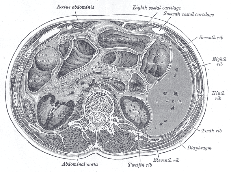 Transverse section through the middle of the first lumbar vertebra, showing the relations of the pancreas.
 -
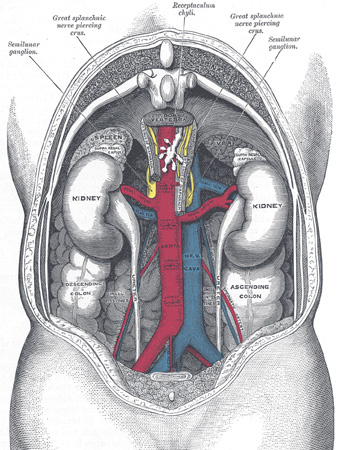
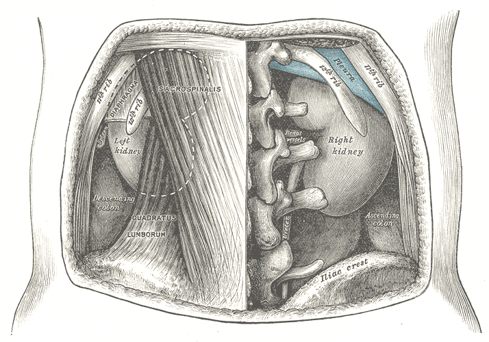 The relations of the kidneys from behind.
 -
-
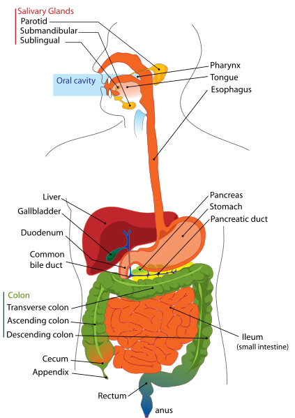 Digestive system


External links
External links
- Template:SUNYAnatomyFigs – “The large intestine.”
- Template:SUNYAnatomyLabs
Looking for the patient version?
© 2026 MyEClinic – IFTM Institut für Telematik in der Medizin GmbH