

Health Dictionary
Reliable, easy-to-read patient information on common medical conditions — written in plain language, free of jargon, and grounded in current evidence. Browse the most frequent diagnoses, understand what they mean for everyday life, and learn which steps and questions help you have a focused conversation with your doctor.
Electrocardiographic Findings in LVH
Introduction
Synonyms and keywords: LVH; LVH with strain; strain pattern
Overview
Overview
Left ventricular hypertrophy is associated with increased QRS voltage on the EKG and a strain pattern or inverted checkmark pattern to the T wave in the lateral leads. There are a variety of criteria used to diagnose left ventricular hypertrophy (LVH) on the EKG.
Diagnosis
Diagnosis
Electrocardiography
Sokolow and Lyon Criteria
- Add the depth of the S wave in V1 to the height of the R wave in lead V5 or V6 (whichever is taller) and if the sum is greater than 35 mm then LVH is present.
- This criterion correlates well with the thickness of the LV walls and the diameter of the LV cavity as determined by ECHO.
- Sensitivity 22% and specificity of 100%.[1]
Effects of Left Anterior Hemiblock on Diagnosing Acute myocardial infarction and Left Ventricular Hypertrophy
LAHB may be a cause of poor R wave progression across the precordium causing a pseudoinfarction pattern mimicking an anteroseptal infarction. It also makes the electrocardiographic diagnosis of LVH more complicated, because both may cause a large R wave in lead aVL. Therefore to call LVH on an EKG in the setting of an LAHB you should see the presence of a “strain” pattern when you are relying on limb lead criteria to diagnose LVH.[2]
Cornell Voltage Criteria
- Add the height of the R wave in lead aVL to the depth of the S wave in lead V3.
- LVH if the sum is > 28mm in men or > 20 mm in women.
- Sensitivity of 42% and specificity of 96%.[3]
Roberts Criteria
Estes Criteria
- R or S in limb lead: 20 mm or more
- S in V1, V2, or V3: 25 mm or more 3 points
- R in V4, V5, or V6: 25 mm or more
- Any ST shift (without digitalis): 3 points
- Typical “strain” ST T (with digitalis): 1 points
- LAD: 15 degrees or more: 2 points
- QRS interval: 0.09 seconds or more: 1 point
- Intrinsicoid deflection in V5 or V6 of 0.04 seconds or more: 1 point
- P terminal force in V1 more than 0.04 sec: 1 point
Total possible: 13 points
Total of 5 points = LVH, 4 points = probable LVH[5]
Electrocardiographic Examples
-
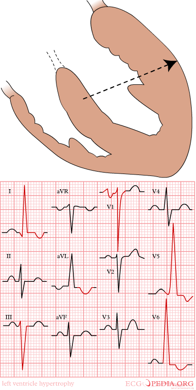 Mechanism of left ventricular hypertrophy

-
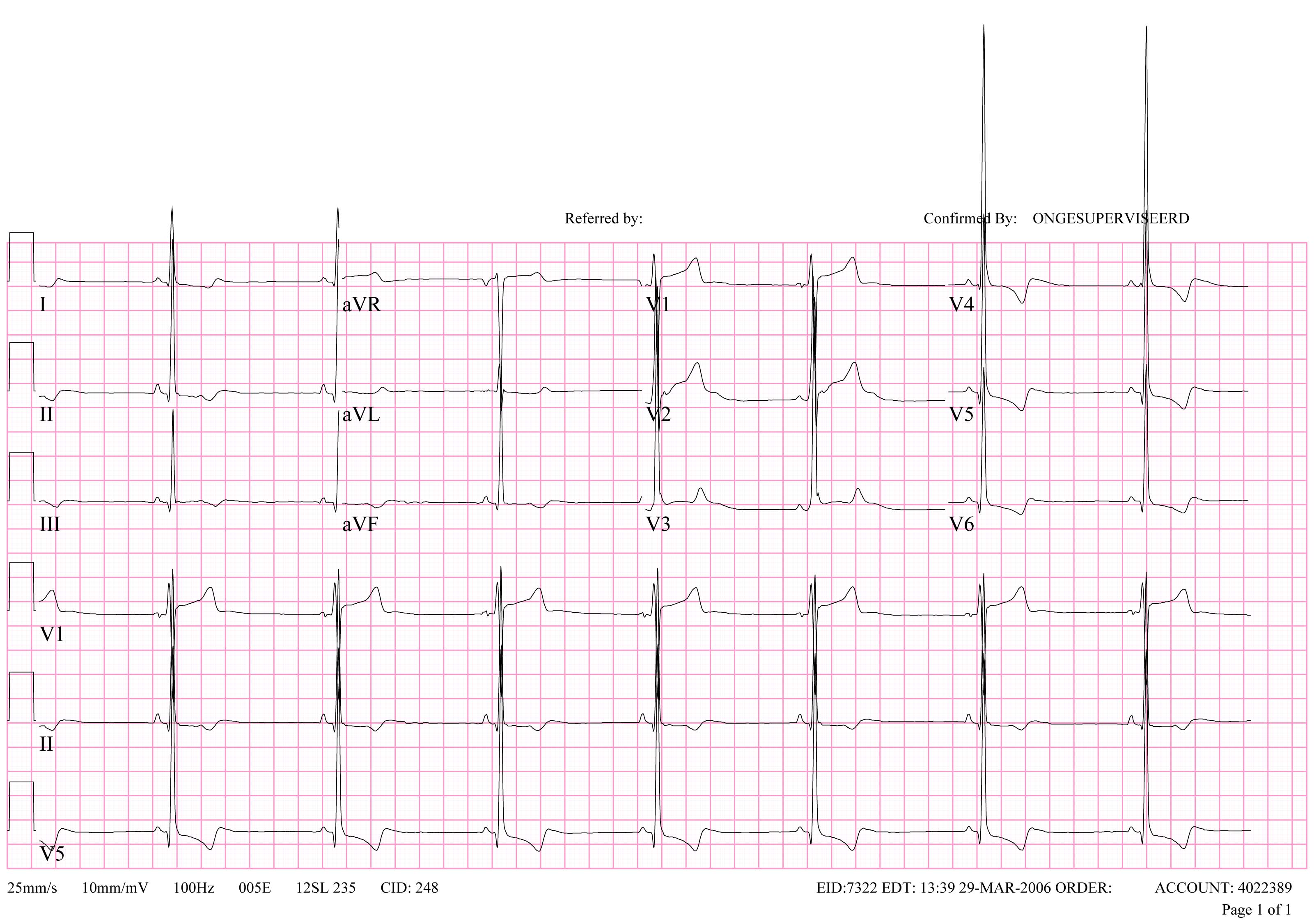
-
Extreme left ventricular hypertrophy in a patient with severe aortic valve stenosis

-
LVH in subendocardial ischemia with positive cardiovascular markers
-
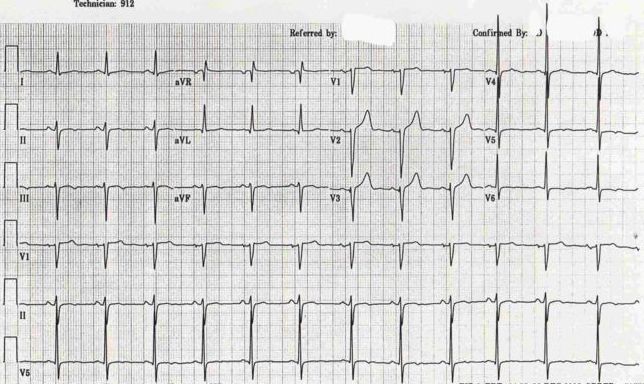 LVH + Left Anterior Hemiblock

References
References
- ↑ Sokolow, M, and Lyon, T.P.: The Ventricular Complex As Obtained By Unipolar Limb Leads. Am. Heart J. 1949:37,161.
- ↑ Hammill S. C. Electrocardiographic diagnoses: Criteria and definitions of abnormalities, Chapter 18, MAYO Clinic, Concise Textbook of Cardiology, 3rd edition, 2007 ISBN 0-8493-9057-5
- ↑ Casale, P., Electrocardiographic detection of left ventricular hypertrophy: Development and prospective evaluation of improved criteria. J. Am. Coll Cardiol. 1985:6,572
- ↑ Roberts, W. and Podalak, M: The king of hearts: Analysis of 23 patients with hearts weighing 1,000 grams or more. Am J. Cardiol. 1985:55,485.
- ↑ Surawicz, B.: Electrocardiographic diagnosis of chamber enlargement. J. Am. Coll. Cardiol. 1986: 8,711.
Editor-In-Chief: C. Michael Gibson, M.S., M.D. [2]; Cafer Zorkun, M.D., Ph.D. [3]