

Health Dictionary
Reliable, easy-to-read patient information on common medical conditions — written in plain language, free of jargon, and grounded in current evidence. Browse the most frequent diagnoses, understand what they mean for everyday life, and learn which steps and questions help you have a focused conversation with your doctor.
Enterobiasis laboratory findings
Introduction
Overview
Overview
Diagnosis of enterobiasis is often made clinically by observing the female worm(s) in the peri-anal region, but can also be made using the “scotch-tape” test, in which the sticky side of a strip of cellophane tape is pressed against the peri-anal skin, then examined under a microscope for pinworm eggs.
Laboratory Findings
Laboratory Findings
Scotch Tape Test
It is also called Hall or National Institute of Health swab. Sticky side of a strip of cellophane tape is pressed against the peri-anal skin, then examined under a microscope for pinworm eggs. The test is repeated for five consecutive mornings to increase the sensitivity to 99%. It is done prior to washing or defecation. The diagnostic characteristics of egg are: size 50-54 µm by 20-27 µm; typical elongated shape, with one convex side and one flattened side and colorless shell.[1][2]
Stool analysis
Stool analysis for ova and parasites is of low diagnostic yield. The actual worms may be seen in the host’s feces; however the eggs are invisible to the naked eye.[1]
Histology
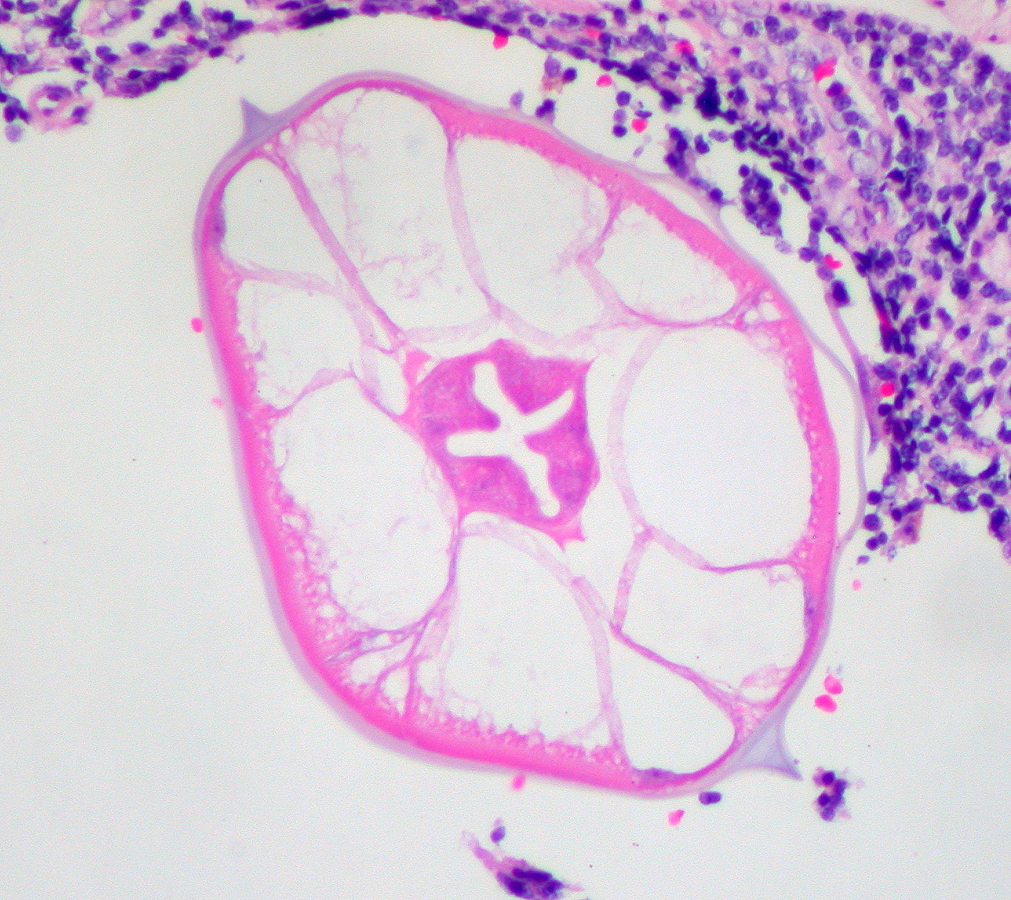
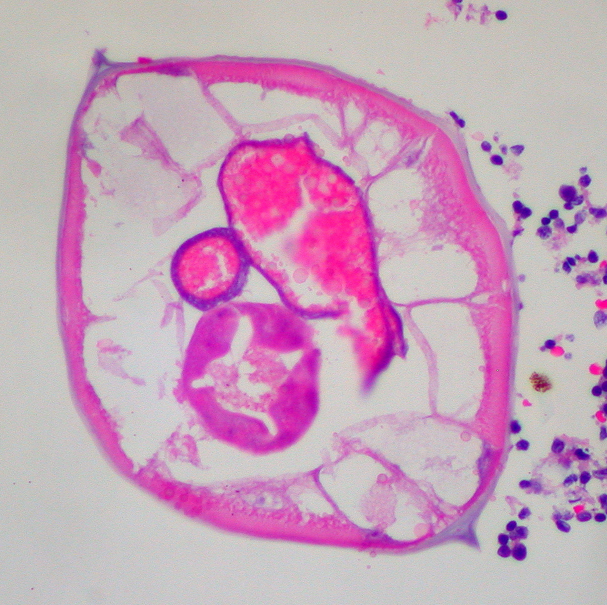
On histologic cross-section alae or wings (running the length of the worm) are identifying features of the pinworm (see micrograph).[3]
Images
-
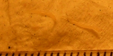 Two pinworms, captured on emergence from the anus. Markings are 1 mm apart.
 -
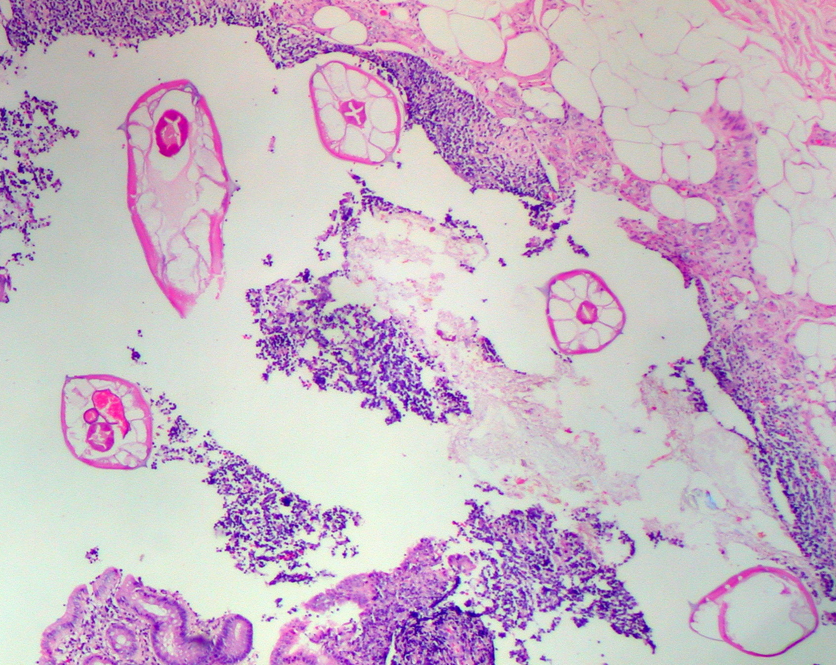 Pinworms are sometimes diagnosed incidentally by pathology. Micrograph of pinworms in the appendix. H&E stain.
 -
-
 High magnification micrograph of a pinworm in cross-section in the appendix. H&E stain.
 -
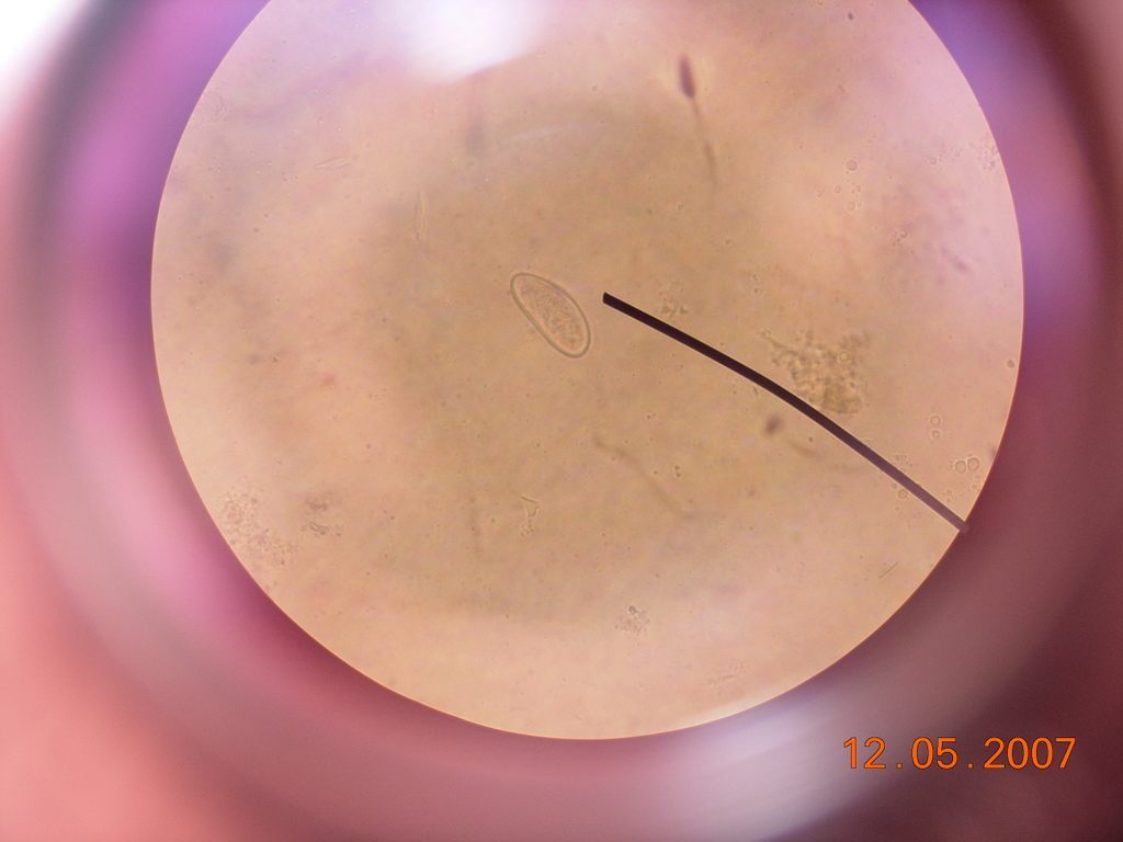 Enterobius vermicularis egg under a light microscope.
 -
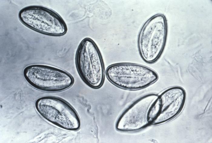 Pinworm eggs are easily seen under a microscope.
 -
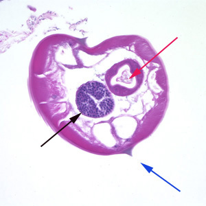 Pinworms are sometimes diagnosed incidentally by pathology. Micrograph of male pinworm in cross-section. Alae (blue arrow), intestine (red arrow) and testis (black arrow). H&E stain.
 -
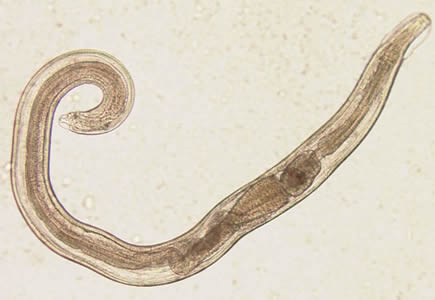 A pinworm (Enterobius vermicularis).


Videos Showing Pinworm Egg Under Microscope
{{#ev:youtube|YNrkWStDdmo}}
References
References
- ↑ 1.0 1.1 Caldwell JP (1982). “Pinworms (enterobius vermicularis)”. Can Fam Physician. 28: 306–9. PMC 2306321. PMID 21286054.
- ↑ Cook GC (1994). “Enterobius vermicularis infection”. Gut. 35 (9): 1159–62. PMC 1375686. PMID 7959218.
- ↑ Diagnostic Findings Enterobiasis. Centers for Disease Control and Prevention. URL:http://www.dpd.cdc.gov/dpdx/HTML/Enterobiasis.htm. Accessed on: August 6, 2008.
Editor-In-Chief: C. Michael Gibson, M.S., M.D. [1] Associate Editor(s)-in-Chief: Furqan M M. M.B.B.S[2]