
Goblet cell
Editor-In-Chief: C. Michael Gibson, M.S., M.D. [1]
Overview
Overview
Goblet cells are glandular simple columnar epithelial cells whose sole function is to secrete mucus. They secrete using both apocrine and merocrine methods of secretion.
The majority of the cell’s cytoplasm is occupied by mucinogen granules, except at the bottom. Rough endoplasmic reticulum, mitochondria, the nucleus, and other organelles are concentrated in the basal portion. The apical plasma membrane projects microvilli to increase surface area for secretion.
Locations
Locations
They are found scattered among the epithelial lining of many organs, especially the intestinal and respiratory tracts. In the respiratory tract, they are found inside the trachea, bronchus, and larger bronchioles.
Histology
Histology
In mucicarmine stains, goblet cells can be easily identified by the deep red mucin found within their cell bodies.
The nuclei of goblet cells tend to be displaced toward the basal end of the cell body, close to basement membrane, leading to intense basophilic staining.
Etymology
Etymology
The term goblet refers to these cells’ goblet-like shape. The apical portion is shaped like a cup, as it is distended by abundant mucinogen granules; its basal portion is shaped like a stem, as it is narrow for lack of these granules.
There are other cells which secrete mucus (as in the fundic glands of the stomach[1]), but they are not usually called “goblet cells” because they do not have this distinctive shape.
Basal secretion
Basal secretion
This is the normal base level secretion of mucus. The continuous secretion is accomplished by cytoskeletal movement of secretory granules.
Stimulated secretion
Stimulated secretion
Secretion may be stimulated by dust, smoke, etc.
Other stimuli include viruses, bacteria, etc.
Additional images
Additional images
-
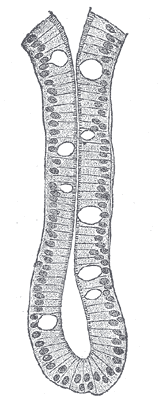 An intestinal gland from the human intestine.
 -
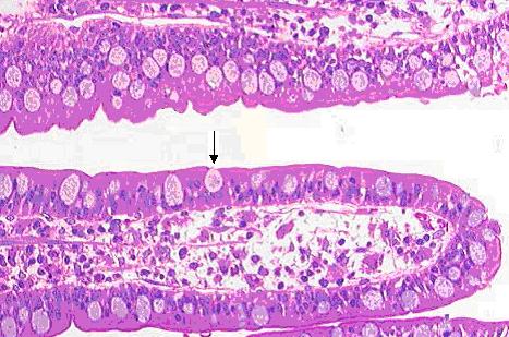 Goblet cell in ileum

References
References
- ↑ Histology image: 11303loa – Histology Learning System at Boston University – Digestive System: Alimentary Canal: fundic stomach, gastric glands, lumen”
External links
External links
- Histology at KUMC epithel-epith08 “Slide 8: Trachea”
- Template:EMedicineDictionary
- Goblet Cells at cvmbs.colostate.edu
- Diagram at uwlax.edu
- Template:BiowebUW
de:Becherzelle
it:Cellula mucipara caliciforme
nl:Slijmbekercel
fi:Pikarisolu
sv:Bägarcell
Looking for the patient version?
© 2026 MyEClinic – IFTM Institut für Telematik in der Medizin GmbH