
Hamartoma MRI
Editor-In-Chief: C. Michael Gibson, M.S., M.D. [1]Associate Editor(s)-in-Chief: Maria Fernanda Villarreal, M.D. [2] Vamsikrishna Gunnam M.B.B.S [3]
Overview
Overview
MRI is the modality of choice for assessment of hypothalamic, spleen, kidney, and other abdominal hamartomas. On MRI, hamartoma is characterized by a heterogeneous signal in T1 and high signal due to fat and cartilaginous components in T2.
MRI
MRI
MRI may be helpful in the diagnosis of hamartomas. Findings on MRI suggestive of hamartomas include:[1][2][3]
- T1: isointense to cerebral cortex
- T1 contrast: no contrast enhancement
- T2: iso to hyperintense to cerebral cortex, the higher the proportion of glial cells, the higher the T2 signal.
- MR spectroscopy
Gallery
Gallery
Hypothalamic Hamartoma
-
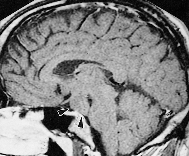 MRI showing hypothalamic hamartoma(Images courtesy of RadsWiki)

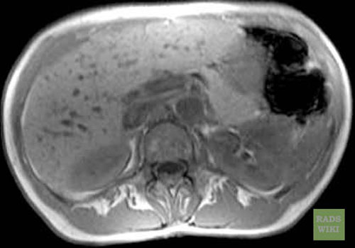
Biliar Hamartomas
-
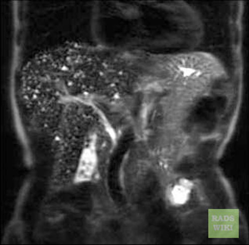 Biliary hamartomas(Images courtesy of RadsWiki)
 -
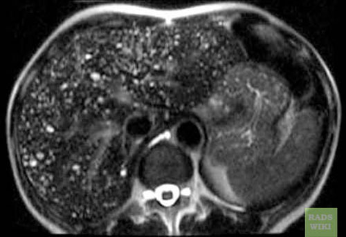 Biliary hamartomas(Images courtesy of RadsWiki)
 -
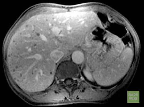 Biliary hamartomas(Images courtesy of RadsWiki)
 -
 Biliary hamartomas(Images courtesy of RadsWiki)


References
References
- ↑ Hypothalamic hamartoma.Dr Donna D’Souza et al. Radiopedia.http://radiopaedia.org/articles/pulmonary-hamartoma-1 Accessed on December 09, 2015
- ↑ Leiter Herrán F, Restrepo CS, Alvarez Gómez DI, Suby-Long T, Ocazionez D, Vargas D (March 2017). “Hamartomas from head to toe: an imaging overview”. Br J Radiol. 90 (1071): 20160607. doi:10.1259/bjr.20160607. PMC 5601532. PMID 27936889.
- ↑ Saleem SN, Said AH, Lee DH (2007). “Lesions of the hypothalamus: MR imaging diagnostic features”. Radiographics. 27 (4): 1087–108. doi:10.1148/rg.274065123. PMID 17620469.
Looking for the patient version?
© 2026 MyEClinic – IFTM Institut für Telematik in der Medizin GmbH