

Health Dictionary
Reliable, easy-to-read patient information on common medical conditions — written in plain language, free of jargon, and grounded in current evidence. Browse the most frequent diagnoses, understand what they mean for everyday life, and learn which steps and questions help you have a focused conversation with your doctor.
Hepatic veins
Introduction
Overview
Overview
In human anatomy, the hepatic veins are the blood vessels that drain de-oxygenated blood from the liver and blood cleaned by the liver (from the stomach, pancreas, small intestine and colon) into the inferior vena cava.
They arise from the substance of the liver, more specifically the central vein of the liver lobule.
None of the hepatic veins have valves.
Groups
Groups
They can be differentiated into two groups, the upper group and lower group.
- The upper group typically arises from the posterior aspect of the liver, are three in number, and drain the quadrate lobe and left lobe.
- The lower group arise from the right lobe and caudate lobe, are variable in number, and are typically smaller than those in the upper group.
External links
External links
- Hepatic Histology: The Lobule – Describes the liver lobule and central vein.
- Hepatic veins – definition – medterms.com
- Template:EMedicineDictionary
Images of the hepatic veins
- Hepatic veins – Ultrasound – University of the Health Sciences in Bethesda, Maryland
- 3-D reconstruction of the liver anatomy (for transplantation) – MeVis Distant Services
- Hepatic veins – CT angiogram – Contrast Techniques for Hepatic Multidetector CT Angiography – Havard Medical School.
- Template:ViennaCrossSection
Additional images
Additional images
-
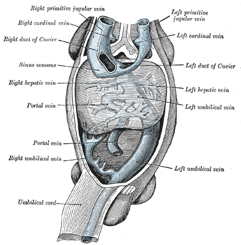 Human embryo with heart and anterior body-wall removed to show the sinus venosus and its tributaries.
 -
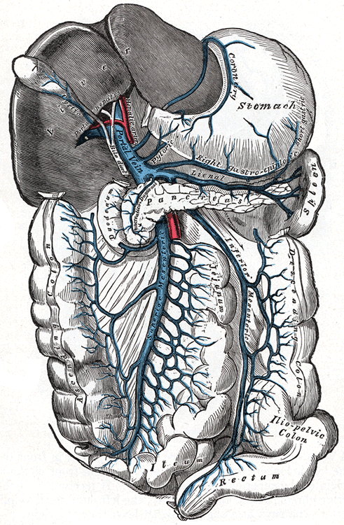 The portal vein and its tributaries.
 -
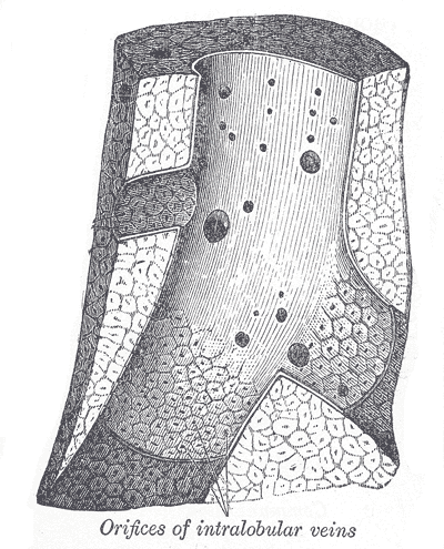 Longitudinal section of a hepatic vein.


Editor-In-Chief: C. Michael Gibson, M.S., M.D. [1]