
Hip examination
Editor-In-Chief: C. Michael Gibson, M.S., M.D. [1]
In medicine, the hip examination, or hip exam, is undertaken when a patient has a complaint of hip pain and/or signs and/or symptoms suggestive of hip joint pathology. It is a physical examination maneuver.
The hip examination, like all examinations of the joints, is typically divided into the following sections:
- Position/lighting/draping
- Inspection
- Palpation
- Motion
- Special maneuvers
The middle three steps are often remembered with the saying look, feel, move.
Position/Lighting/Draping
Position/Lighting/Draping
Position – for most of the exam the patient should be supine and the bed or examination table should be flat. The patient’s hands should remain at her sides with her head resting on a pillow. The knees and hips should be in the anatomical position (knee extended, hip neither flexed nor extended).
Lighting – adjusted so that it is ideal.
Draping – both of the patient’s hips should be exposed so that the quadriceps muscles and greater trochanter can be assessed.
Inspection
Inspection
Inspection done while the patient is standing
The hip should be examined for:
- Abnormal gait – i.e. antalgic gait
Inspection done while supine
The hip should be examined for:
- Masses
- Scars
- Lesions
- Signs of trauma/previous surgery
- Bony Alignment (rotation, leg length)
- Muscle bunk and symmetry at the hip and knee
Measures
- True leg length – anterior superior iliac spine (ASIS) to medial malleolus
- Apparent leg length – umbilicus to medial malleolus
In hip fractures the affected leg is often shortened and externally rotated.
Palpation
Palpation
The hip joint lies is deep and cannot normally be directly palpated.
To assess for pelvic fracture one should palpate the:
- Ischial spines
- Pubic rami
Movement
Movement
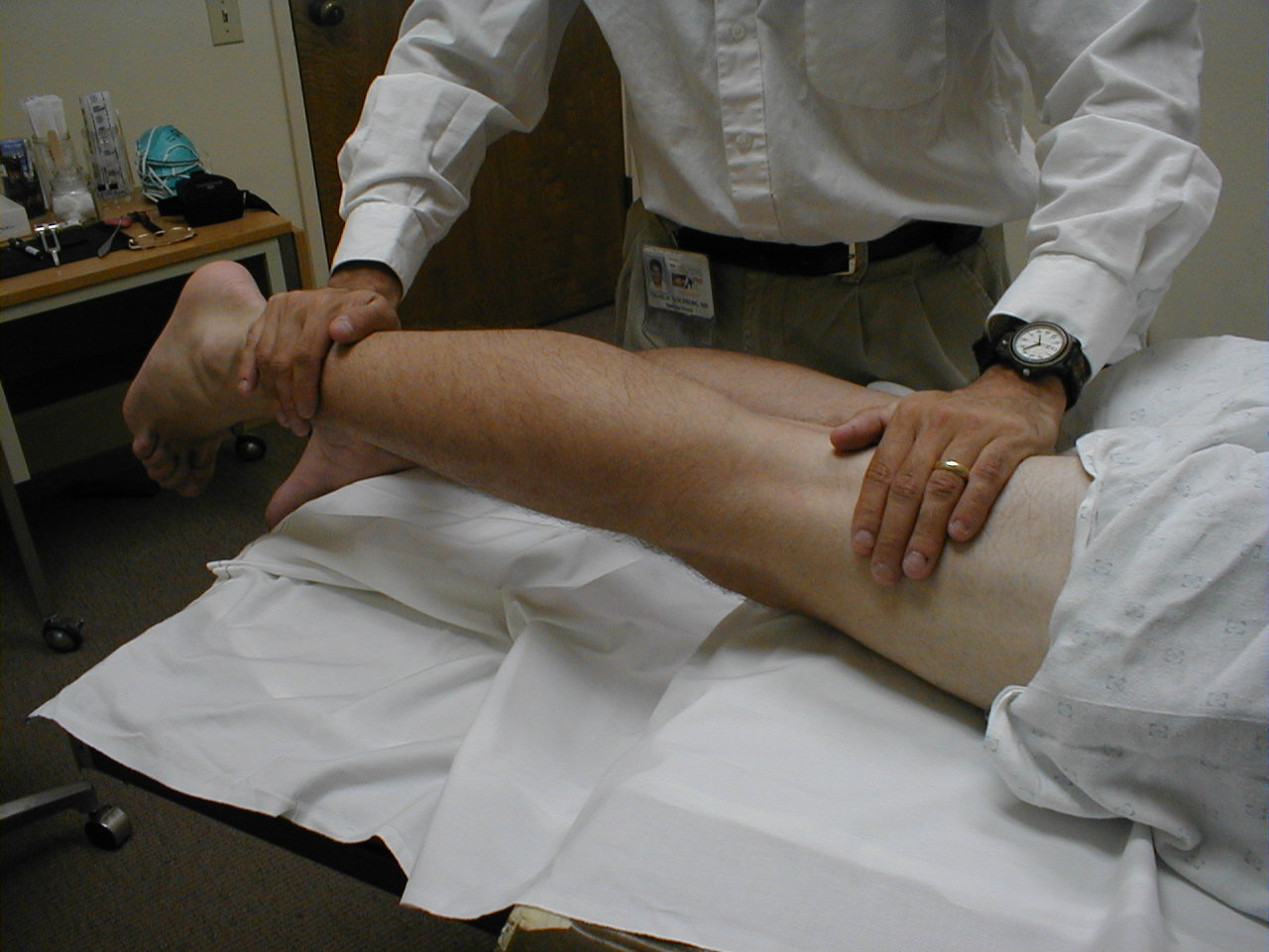
- Internal rotation – with knee and hip both flexed at 90 degrees the ankle is abducted.
- External rotation – with knee and hip both flexed at 90 degrees the ankel is adducted.
- Flexion
- Extension – done with the patient on their side. Alignment should be assessed by palpation of the ASIS, PSIS and greater trochanter.
- Abduction – assessed whilst palplating the contralateral ASIS.
- Adduction – assessed whilst palpating the ipsilateral ASIS.
- Assessment for a hidden flexion contracture of the hip – hip flexion contractures may be occult, due to compensation by the back. They are assessed by:
- Placing a hand behind the lumbar region of back
- Getting the patient to fully flex the contralateral hip.
- The hand in the lumbar region is used to confirm the back is straightened (flexed relative to the anatomic position). If there is a flexion contracture in the ipsilateral hip it should evident, as the hip will appear flexed.
Normal range of motion
- Internal rotation – 35°
- External rotation – 45°
- Flexion – 135° (L 2, 3, 4)
With the patient seated, place your hand on top of one thigh and instruct the patient to lift the leg up from the table. The main hip flexor is the Iliopsoas muscle, innervated by the femoral nerve.
(Image courtesy of Charlie Goldberg, M.D., UCSD School of Medicine and VA Medical Center, San Diego, California)
-
 Hip flexion

- Extension – 25° (L5, S1):
With the patient lying prone, direct the patient to lift their leg off the table against resistance. Test each leg separately. The main hip extensor is the gluteus maximus, innervated by inferior gluteal nerve.
(Image courtesy of Charlie Goldberg, M.D., UCSD School of Medicine and VA Medical Center, San Diego, California)
-
 Hip extension

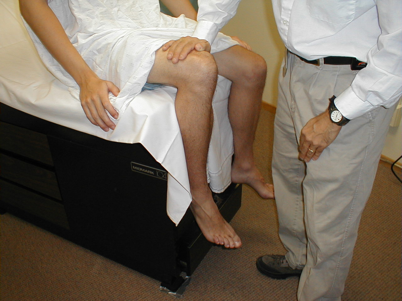
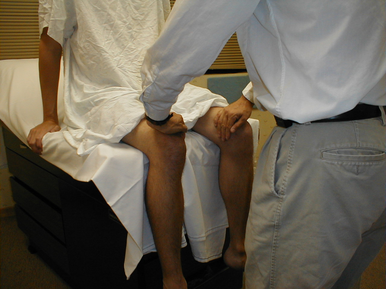
- Abduction – 45° (L 4, 5, S1):
Place your hands on the outside of either thigh and direct the patient to separate their legs against resistance. This movement is mediated by a number of muscles.
(Image courtesy of Charlie Goldberg, M.D., UCSD School of Medicine and VA Medical Center, San Diego, California)
-
 Hip abduction

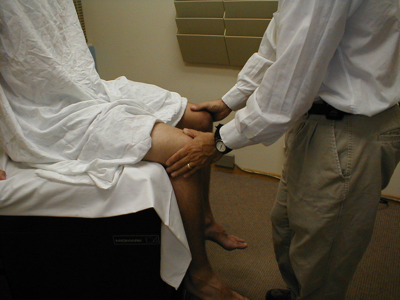
- Adduction – 25° (L 2, 3, 4):
Place your hands on the inner aspects of the thighs and repeat the maneuver. A number of muscles are responsible for adduction. They are innervated by the obturator nerve.
(Image courtesy of Charlie Goldberg, M.D., UCSD School of Medicine and VA Medical Center, San Diego, California)
-
 Hip adduction

Video: Hip Examination
{{#ev:youtube|5LNYdJIrWYo}}
Special maneuvers
Special maneuvers
- Trendelenburg’s test: Telescoping axial movement is tested with knee bent 90 degrees and lying on couch. Tests for dislocation
Other tests
Other tests
A knee examination should be undertaken in the ipsilateral knee to rule-out knee pathology.
Looking for the patient version?
© 2026 MyEClinic – IFTM Institut für Telematik in der Medizin GmbH