
Horner's syndrome physical examination
Editor-In-Chief: C. Michael Gibson, M.S., M.D. [1]
Please help WikiDoc by adding more content here. It’s easy! Click here to learn about editing.
Overview
Overview
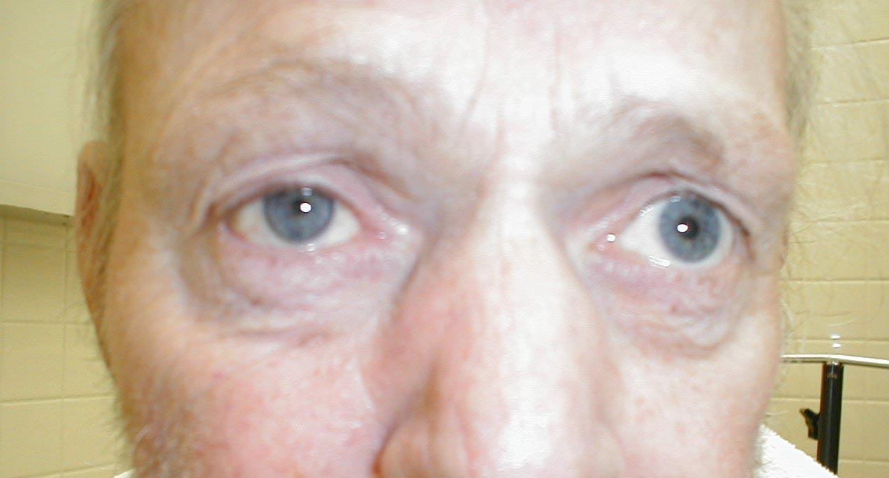
An eye examination may show changes in how the pupil opens or closes and eyelid drooping. A complete medical and nervous system (neurological) examination can show whether any other parts of the body are affected.
Physical Examination
Physical Examination
Signs found in all patients on affected side of face include ptosis (drooping upper eyelid from loss of sympathetic innervation to the Müller– Rouget muscle), upside-down ptosis (slight elevation of the lower lid), and miosis (constricted pupil) and dilation lag. Enophthalmos (the impression that the eye is sunk in) and anhidrosis (decreased sweating) on the affected side of the face, loss of ciliospinal reflex and blood shot conjunctiva may occur depending on the site of lesion.
In children Horner’s syndrome sometimes leads to a difference in eye color between the two eyes (heterochromia).[1] This happens because a lack of sympathetic stimulation in childhood interferes with melanin pigmentation of the melanocytes in the superficial stroma of the iris.
-
 Horner’s Syndrome

References
References
- ↑ Gesundheit B, Greenberg M (2005). “Medical mystery: brown eye and blue eye–the answer”. N Engl J Med. 353 (22): 2409–10. PMID 16319395.
Looking for the patient version?
© 2026 MyEClinic – IFTM Institut für Telematik in der Medizin GmbH