

Health Dictionary
Reliable, easy-to-read patient information on common medical conditions — written in plain language, free of jargon, and grounded in current evidence. Browse the most frequent diagnoses, understand what they mean for everyday life, and learn which steps and questions help you have a focused conversation with your doctor.
Hydrocephalus CT
Introduction
Overview
Overview
Ventricular enlargement not entirely attributable to cerebral atrophy or congenital enlargement (Evans index >0.3). No macroscopic obstruction to CSF flow. Enlargement of the temporal horns of the lateral ventricles not entirely attributable to hippocampus atrophy. Callosal angle of 40º or greater. Evidence of altered brain water content, including periventricular signal changes on CT and MRI not attributable to microvascular ischemic changes or demyelination. An aqueductal or fourth ventricular flow void.
CT
CT
- The Ct scan findings are given below:[1][2][3]
- Ventricular enlargement not entirely attributable to cerebral atrophy or congenital enlargement (Evans index >0.3).
- No macroscopic obstruction to CSF flow.
- At least one of the following supportive features:
- Enlargement of the temporal horns of the lateral ventricles not entirely attributable to hippocampus atrophy;
- Callosal angle of 40º or greater;
- Evidence of altered brain water content, including periventricular signal changes on CT and MRI not attributable to microvascular ischemic changes or demyelination;
- An aqueductal or fourth ventricular flow void.


Hydrocephalus
-
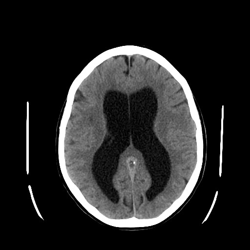 Hydrocephalus
 -
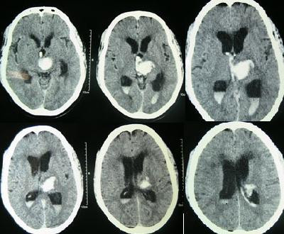 Intracerebral hemorrhage
 -
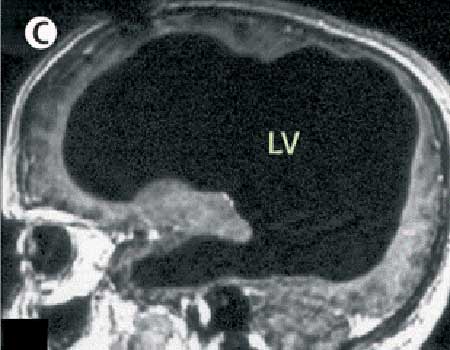 DWS: All of the black in the middle is cerebrospinal fluid and the brain matter is the rim of white along the outside of the skull.


References
References
- ↑ Damasceno BP (2015). “Neuroimaging in normal pressure hydrocephalus”. Dement Neuropsychol. 9 (4): 350–355. doi:10.1590/1980-57642015DN94000350. PMC 5619317. PMID 29213984.
- ↑ Toma AK, Holl E, Kitchen ND, Watkins LD (April 2011). “Evans’ index revisited: the need for an alternative in normal pressure hydrocephalus”. Neurosurgery. 68 (4): 939–44. doi:10.1227/NEU.0b013e318208f5e0. PMID 21221031.
- ↑ Mori E, Ishikawa M, Kato T, Kazui H, Miyake H, Miyajima M, Nakajima M, Hashimoto M, Kuriyama N, Tokuda T, Ishii K, Kaijima M, Hirata Y, Saito M, Arai H (2012). “Guidelines for management of idiopathic normal pressure hydrocephalus: second edition”. Neurol. Med. Chir. (Tokyo). 52 (11): 775–809. PMID 23183074.
Editor-In-Chief: C. Michael Gibson, M.S., M.D. [1]; Associate Editor(s)-in-Chief: Syed Ahsan Hussain, M.D.[2]