

Health Dictionary
Reliable, easy-to-read patient information on common medical conditions — written in plain language, free of jargon, and grounded in current evidence. Browse the most frequent diagnoses, understand what they mean for everyday life, and learn which steps and questions help you have a focused conversation with your doctor.
Internal pterygoid muscle
Introduction
Overview
Overview
The medial pterygoid (or internal pterygoid muscle), is a thick, quadrilateral muscle of mastication.
The mandibular branch of the fifth cranial nerve, the trigeminal nerve, innervates the medial pterygoid muscle.
Origin and insertion
Origin and insertion
It consists of two heads.
- The bulk of the muscle arises as a deep head from the medial surface of the lateral pterygoid plate.
- The smaller, superficial head originates from the maxillary tuberosity and the pyramidal process of the palatine bone.
Its fibers pass downward, lateral, and posterior, and are inserted, by a strong tendinous lamina, into the lower and back part of the medial surface of the ramus and angle of the mandible, as high as the mandibular foramen. The insertion joins the masseter muscle to form a common tendinous sling which allows the medial pterygoid and masseter to be powerful elevators of the jaw.
As stated by one Dr. Anderson of UDMSD, the medial pterygoid goes from the canoe to the rough water. (The canoe is the depression created by the medial and lateral plates of pterygoid process, and rough water is the rough attachment surface on the medial aspect of the angle of the mandible.)
Innervation
Innervation
Like the lateral pterygoid, and all other muscles of mastication, the medial pterygoid is innervated by the mandibular branch of the trigeminal nerve (V3).
Actions
Actions
It closes the jaw and help in mastication along with lateral pterygoid in side to side movement of jaw and protrusion. It elevates the jaw, and in some aspect pulls it forward.
Also it elevates mandible.
Additional images
Additional images
-
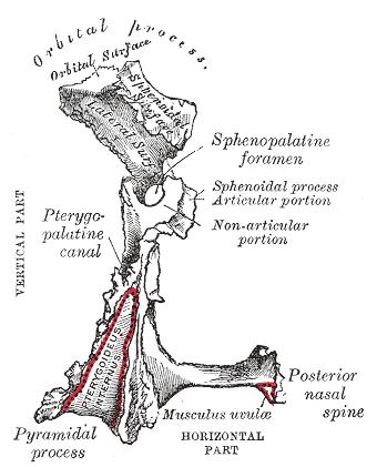 Left palatine bone. Posterior aspect. Enlarged.
 -
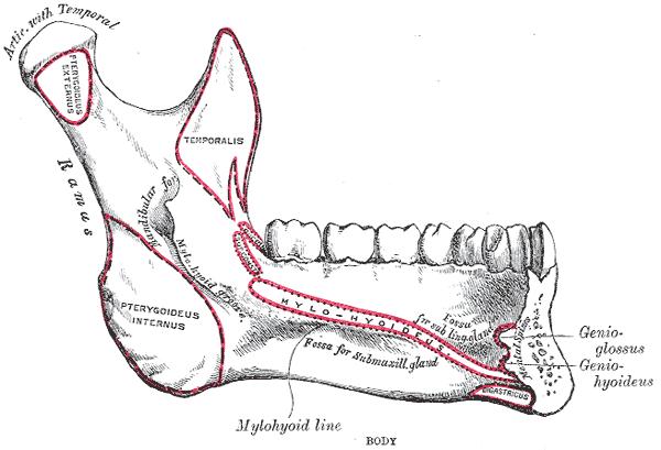 Mandible. Inner surface. Side view.
 -
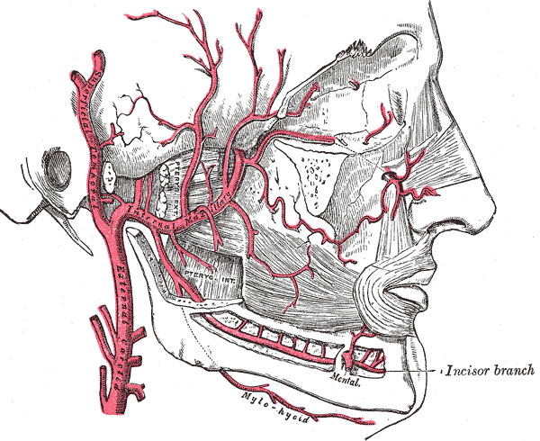 Plan of branches of internal maxillary artery.
 -
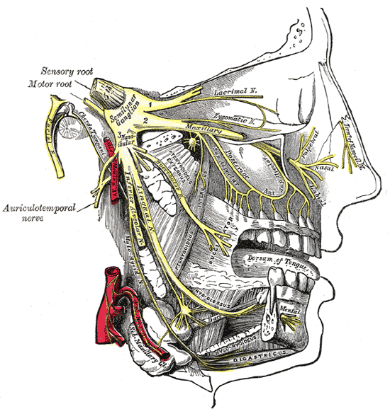 Distribution of the maxillary and mandibular nerves, and the submaxillary ganglion.
 -
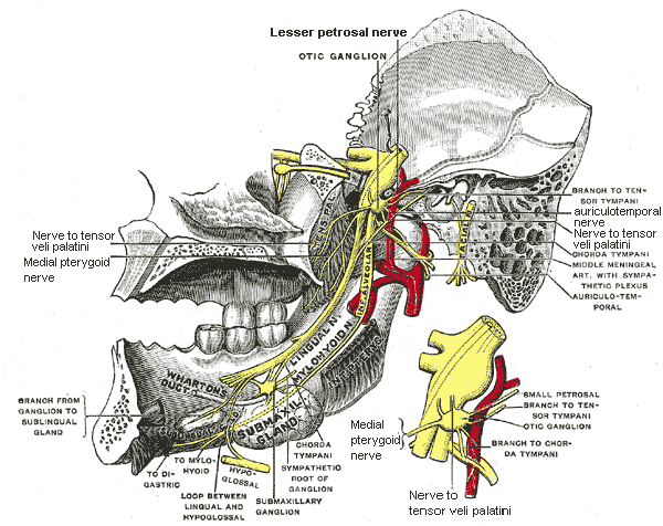 Mandibular division of trifacial nerve, seen from the middle line.
 -
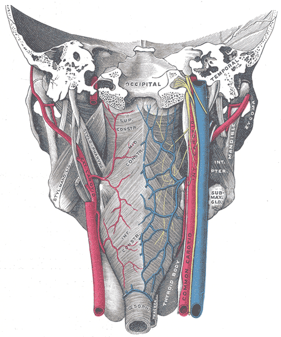 Muscles of the pharynx, viewed from behind, together with the associated vessels and nerves.


Editor-In-Chief: Template:Muscle infobox C. Michael Gibson, M.S., M.D. [1]