

Health Dictionary
Reliable, easy-to-read patient information on common medical conditions — written in plain language, free of jargon, and grounded in current evidence. Browse the most frequent diagnoses, understand what they mean for everyday life, and learn which steps and questions help you have a focused conversation with your doctor.
Intracerebral hemorrhage MRI
Introduction
Overview
Overview
MRI is better than CT for detection of acute and chronic hemorrhage. Therefore it should be the preferred test for accurate diagnosis of patients with suspected intracerebral hemorrhage.[1]
MRI
MRI
MRI is better than CT for detection of acute and chronic hemorrhage. Therefore it should be the preferred test for accurate diagnosis of patients with suspected intracerebral hemorrhage.[1]
- T2 susceptibility-weighted MRI are as sensitive as CT for detection of acute blood and are more sensitive for identification of prior hemorrhage.[1]
- The appearance of Intracerebral hemorrhage on MRI depends on the age of the hematoma and the type of MR sequence (T1-MRI or T2-MRI).
- As the hematoma ages, hemoglobin (Hb) goes through the following stages:[2]
| Stage | Age | Hemoglobin | 1-MRI | 2-MRI |
|---|---|---|---|---|
| Hyperacute | < 24 hours | Intracellular oxy-Hb | Isointense | Very hyperintense |
| Acute | 1-3 days | Intracellular deoxy-Hb | Slightly hypointense | Very hypointense |
| Early subacute | > 3 days | Intracellular met-Hb | Very hyperintense | Very hypointense |
| Late subacute | > 7 days | Extracellular met-Hb | Very hyperintense | Very hypointense |
| Chronic center | > 14 days | Extracellular hemichromes | Isointense | Very hyperintense |
| Chronic rim | > 14 days | Intracellular hemosiderin | Slightly hypointense | Very hypointense |
Microbleeds
Cerebral microbleeds are best visualized on T2* weighted GRE sequences as punctate areas of low signal.[3][4]
Images
Images
The following are images associated with intracerebral hemorrhage involves the left lentiform nucleus and internal capsule.[5]
-
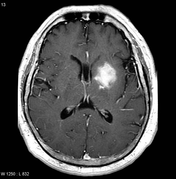 Axial T1
 -
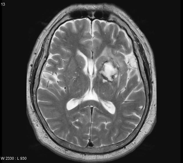 T2
T2 -
 Sagittal T1 C+


References
References
- ↑ 1.0 1.1 1.2 Chalela JA, Kidwell CS, Nentwich LM, Luby M, Butman JA, Demchuk AM; et al. (2007). “Magnetic resonance imaging and computed tomography in emergency assessment of patients with suspected acute stroke: a prospective comparison”. Lancet. 369 (9558): 293–8. doi:10.1016/S0140-6736(07)60151-2. PMC 1859855. PMID 17258669.
- ↑ Bradley WG (1993). “MR appearance of hemorrhage in the brain”. Radiology. 189 (1): 15–26. doi:10.1148/radiology.189.1.8372185. PMID 8372185.
- ↑ Altmann-Schneider I, Trompet S, de Craen AJ, van Es AC, Jukema JW, Stott DJ; et al. (2011). “Cerebral microbleeds are predictive of mortality in the elderly”. Stroke. 42 (3): 638–44. doi:10.1161/STROKEAHA.110.595611. PMID 21233474.
- ↑ Liu W, Liu R, Sun W, Peng Q, Zhang W, Xu E; et al. (2012). “Different impacts of blood pressure variability on the progression of cerebral microbleeds and white matter lesions”. Stroke. 43 (11): 2916–22. doi:10.1161/STROKEAHA.112.658369. PMID 22949472.
- ↑ Intracerebral Hemotrrhage https://radiopaedia.org/cases/intracerebral-haemorrhage-2 Accessed on November 9, 2016
Editor-In-Chief: C. Michael Gibson, M.S., M.D. [1]; Associate Editor(s)-in-Chief: Sara Mehrsefat, M.D. [2]