
Langerhans cell histiocytosis CT
Editor-In-Chief: C. Michael Gibson, M.S., M.D. [1] Associate Editor(s)-in-Chief: Haytham Allaham, M.D. [2]
Overview
Overview
CT scan may be helpful in the diagnosis of Langerhans cell histiocytosis. Findings on CT scan suggestive of Langerhans cell histiocytosis include multiple osteolytic lesions causing full thickness bone destruction.
CT Scan
CT Scan
- Head CT scan may be helpful in the diagnosis of Langerhans cell histiocytosis.[1][2]
- Findings on head CT scan suggestive of Langerhans cell histiocytosis include:[3][4]
- Multiple osteolytic lesions
- Full thickness bone destruction
- “Button sequestrum” sign
Gallery
Gallery
-
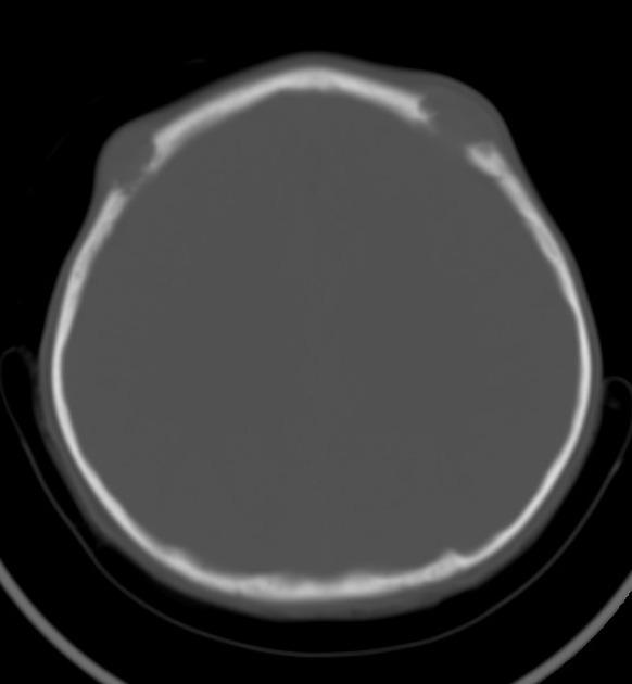 Head CT scan illustrating multiple osteolytic lesions of Langerhans cell histiocytosis
 -
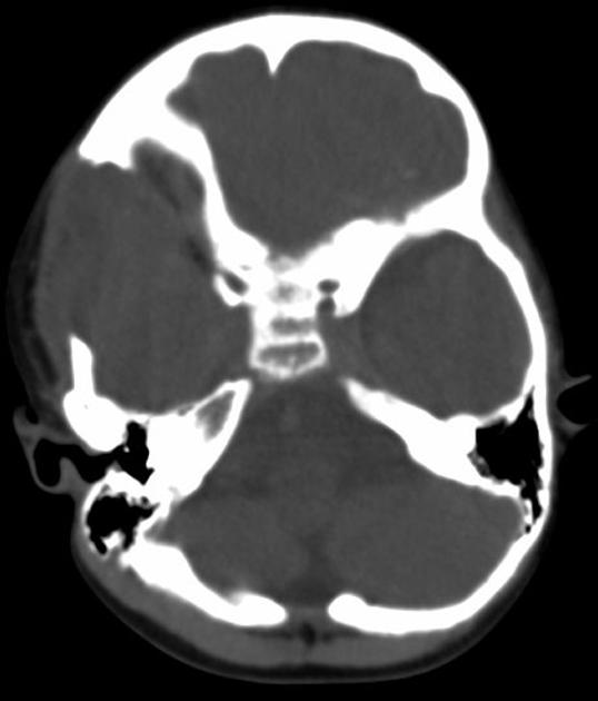 Head CT scan illustrating multiple osteolytic lesions of Langerhans cell histiocytosis
 -
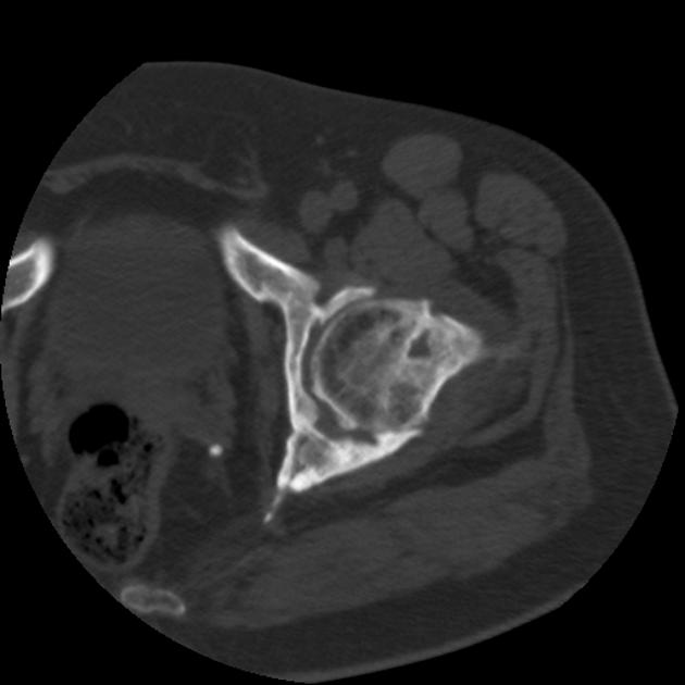 Axial CT scan illustrating full thickness bone destruction of Langerhans cell histiocytosis


References
References
- ↑ Langerhans cell histiocytosis. Radiopeadia (2015) http://radiopaedia.org/articles/langerhans-cell-histiocytosis Accessed on February, 3 2016
- ↑ Khung S, Budzik JF, Amzallag-Bellenger E, Lambilliote A, Soto Ares G, Cotten A; et al. (2013). “Skeletal involvement in Langerhans cell histiocytosis”. Insights Imaging. 4 (5): 569–79. doi:10.1007/s13244-013-0271-7. PMC 3781243. PMID 23907805.
- ↑ Haupt R, Minkov M, Astigarraga I, Schäfer E, Nanduri V, Jubran R, Egeler RM, Janka G, Micic D, Rodriguez-Galindo C, Van Gool S, Visser J, Weitzman S, Donadieu J (February 2013). “Langerhans cell histiocytosis (LCH): guidelines for diagnosis, clinical work-up, and treatment for patients till the age of 18 years”. Pediatr Blood Cancer. 60 (2): 175–84. doi:10.1002/pbc.24367. PMC 4557042. PMID 23109216.
- ↑ Hermans R, De Foer B, Smet MH, Leysen J, Feenstra L, Fossion E, Baert AL (1994). “Eosinophilic granuloma of the head and neck: CT and MRI features in three cases”. Pediatr Radiol. 24 (1): 33–6. PMID 8008491.
Looking for the patient version?
© 2026 MyEClinic – IFTM Institut für Telematik in der Medizin GmbH