
Left atrial enlargement chest x-ray
Editor-In-Chief: C. Michael Gibson, M.S., M.D. [1]; Associate Editor(s)-In-Chief: Cafer Zorkun, M.D., Ph.D. [2]; Varun Kumar, M.B.B.S. [3]
Chest X-Ray
Chest X-Ray
Chest x-ray findings of left atrial enlargement are:
- Double density sign: Occur when the right side of the left atrium pushes behind the right atrial border, appearing as a double density. If large enough it can actually reach beyond the border of the right atrium.
- Convex left atria appendage: usually reflect prior rheumatic heart disease
- Splaying of the carina
- Posterior displacement of the left main stem bronchus on lateral radiograph
- Superior displacement of the left main stem bronchus on frontal view
- Posterior displacement of a barium filled oesophagus or nasogastric tube
Images shown below are courtesy of Radiopedia.com.
-
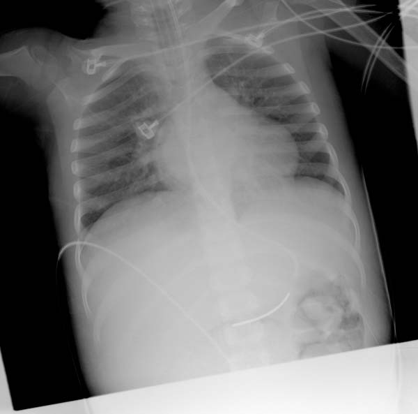 Double density sign
 -
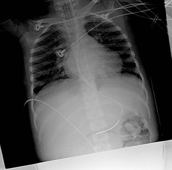 Same patient & the same image. Double density sign. Image’s modified for more contrast and better visualization.
 -
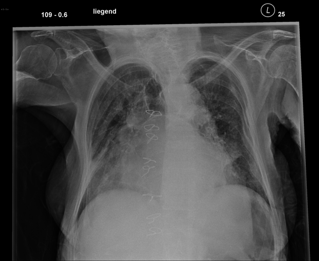 Aside from the dirty lung due to emphysema and pneumonic infiltration in the lower right field you can notice a marked enlargement of the left atrium with splaying of the carina.


Looking for the patient version?
© 2026 MyEClinic – IFTM Institut für Telematik in der Medizin GmbH