
Corpus luteum
Editor-In-Chief: C. Michael Gibson, M.S., M.D. [1]
Overview
Overview
The corpus luteum (Latin for “yellow body”) (plural corpora lutea) is a temporary endocrine structure in mammals, involved in the production of the progestogens which are needed for the maintenance of a pregnancy.
Development and structure
Development and structure
The corpus luteum develops from an ovarian follicle during the luteal phase of the menstrual cycle or estrous cycle, following the release of a mature ovum (egg) from the follicle during ovulation. The follicle first forms a corpus hemorrhagicum before it becomes a corpus luteum, but the term simply refers to the visible collection of blood left after rupture of the follicle, and has no functional significance. While the oocyte (later the zygote) traverses the Fallopian tube into the uterus, the corpus luteum remains in the ovary.
The corpus luteum is typically very large relative to the size of the ovary; in humans, the size of the structure ranges from under 2 cm to 6 cm in diameter. [1]
Its cells develop from the follicular cells surrounding the ovarian follicle:
| Source | Becomes | Secretion |
| The granulosa cells | the inner granulosa lutein layer | progesterone, estrogen |
| Theca cells | the outer theca lutein layer | progesterone, androgens |
Function
Function
It is essential for establishing and maintaining pregnancy in females.
In the ovary, the corpus luteum secretes estrogens and progesterone, which are steroid hormones responsible for the thickening of the endometrium and its development and maintenance, respectively.
When egg is not fertilized
If the egg is not fertilized, the corpus luteum stops secreting progesterone and decays (after approximately 14 days in humans). It then degenerates into a corpus albicans, which is a mass of fibrous scar tissue.
The uterine lining sloughs off without progesterone and is expelled through the vagina (in humans and some great apes, which go through a menstrual cycle). In an estrus cycle the lining degenerates back to normal size.
When egg is fertilized
If fertilized, however, the embryo secretes the hormone human chorionic gonadotropin (hCG) or a similar hormone in other species.
This hormone signals the corpus luteum to continue progesterone secretion, thereby maintaining the thick lining (endometrium) of the uterus, and providing an area rich in blood vessels in which the zygote(s) can develop. From this point on, the corpus luteum is called the corpus luteum graviditatis.
The introduction of the hormone prostaglandin at this point causes the degeneration of the corpus luteum and the abortion of the fetus. However, in placental animals such as humans the placenta eventually takes over progesterone production and the corpus luteum degrades into a corpus albicans without embryo/fetus loss.
Additional images
Additional images
-
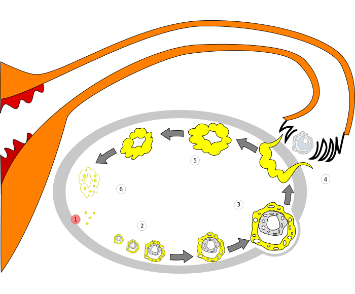 Order of changes in ovary
 -
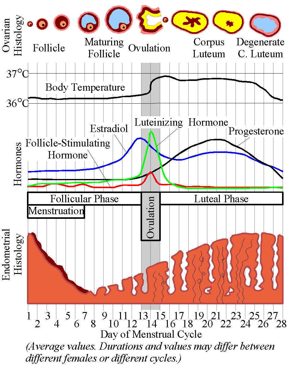 Menstrual Cycle


External links
External links
- Histology image: 18201loa – Histology Learning System at Boston University
- Template:SUNYAnatomyLabs – “The Female Pelvis: The Ovary”
Looking for the patient version?
© 2026 MyEClinic – IFTM Institut für Telematik in der Medizin GmbH